
A New PET Brain Scan Could Bring Us One Step Closer to Diagnosing CTE During Life
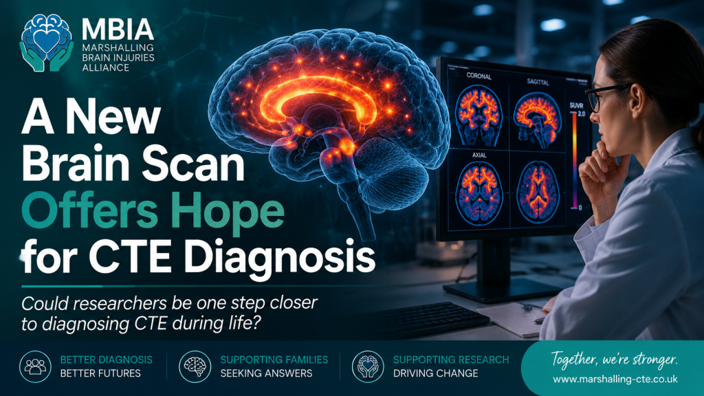
Could one of the biggest challenges in CTE research finally be moving towards a solution?
Researchers have recently announced promising results from an early study investigating a new PET brain scan tracer that may be able to detect the abnormal tau protein associated with Chronic Traumatic Encephalopathy (CTE) in living people.
Although this research is still in its early stages, many experts believe it represents one of the most exciting developments in CTE research for several years.
For people living with persistent symptoms after repeated head impacts—and for the families who support them—it offers something that has often been in short supply:
Hope.
Why Is Diagnosing CTE So Difficult?
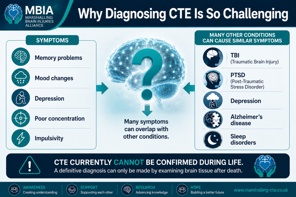
At present, CTE cannot be definitively diagnosed during life.
Doctors can evaluate symptoms, medical history and exposure to repeated head impacts, but a confirmed diagnosis currently requires examination of brain tissue after death.
This creates enormous challenges.
Many people experience symptoms that may be consistent with CTE, including:
- Memory problems
- Difficulty concentrating
- Slower thinking
- Mood changes
- Depression
- Anxiety
- Impulsivity
- Problems controlling emotions
The difficulty is that these symptoms can also occur in many other conditions, including:
- Previous traumatic brain injury
- Post-concussion syndrome
- PTSD
- Depression
- Anxiety disorders
- Alzheimer's disease
- Frontotemporal dementia
- Sleep disorders
This is why researchers around the world are searching for reliable ways to identify CTE during life.
What Is A PET Scan?
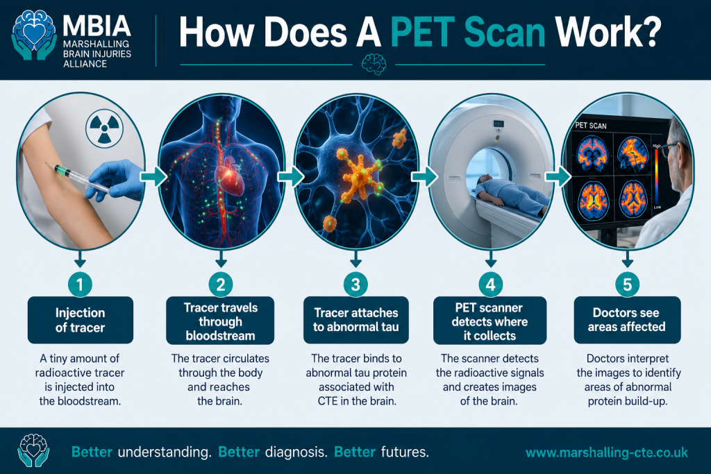
PET stands for Positron Emission Tomography.
Unlike MRI or CT scans, which show the structure of the brain, PET scans can show how certain proteins or chemicals behave inside the brain.
Researchers inject a tiny amount of a radioactive tracer.
The tracer travels through the brain and attaches to a specific target.
The scanner then shows where that tracer has accumulated.
Different tracers can be designed to identify different diseases.
What Did This Study Test?
Researchers investigated a brand-new PET tracer called:
¹⁸F-OXD-2314
This tracer has been specifically designed to attach to the abnormal form of tau protein that accumulates in CTE.
Tau proteins normally help support nerve cells.
However, after years of repeated head impacts, tau can become abnormal, clump together and spread through certain areas of the brain.
These abnormal tau deposits are considered one of the defining features of CTE.
Who Took Part?
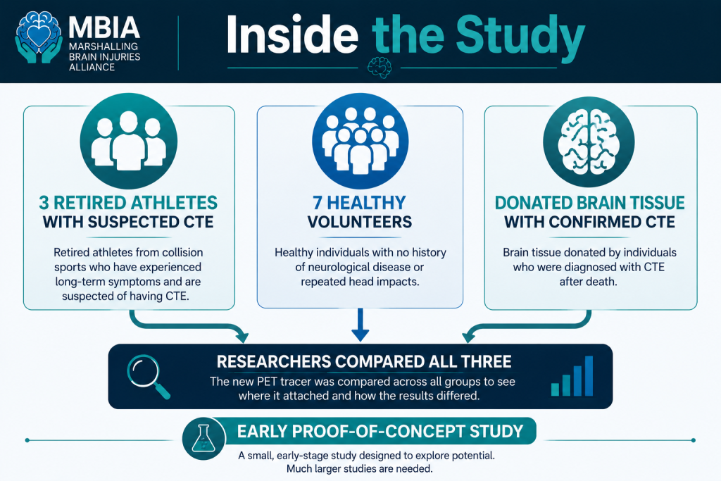
This was a very small proof-of-concept study.
Researchers scanned:
- Three retired collision sport athletes who were suspected of having CTE
- Seven healthy volunteers
The team also tested the tracer on donated brain tissue from people who had been diagnosed with CTE after death.
What Did They Find?
The results were encouraging.
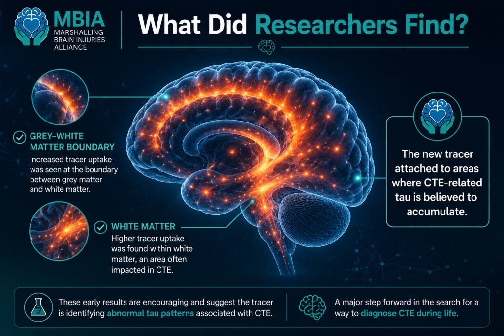
The new tracer appeared to attach to the abnormal tau deposits seen in CTE.
Compared with healthy volunteers, the retired athletes showed:
- Greater tracer uptake in areas where CTE pathology is typically found
- Increased signals at the boundary between grey matter and white matter
- Higher uptake within white matter itself
When researchers tested donated brain tissue, the tracer also successfully bound to confirmed CTE pathology.
These findings suggest the tracer may be identifying the abnormal tau protein researchers hoped it would detect.
Why Is This So Important?
If future research confirms these findings, this technology could eventually help doctors:
🧠 Identify CTE during life
🧠 Distinguish CTE from other neurodegenerative diseases
🧠 Monitor disease progression
🧠 Select suitable patients for future clinical trials
🧠 Test whether new treatments are working
Perhaps most importantly, researchers would no longer have to wait until after someone has died to study the disease.
A Word Of Caution
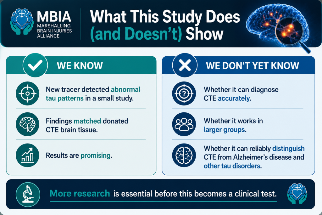
As exciting as these findings are, it is important not to overstate them.
This study included only three people with suspected CTE.
That means we cannot yet say this scan can diagnose CTE.
Much larger studies involving many more participants are still needed.
Researchers must also determine:
- How accurate the tracer is
- Whether it can distinguish CTE from Alzheimer's disease and other tau disorders
- Whether similar findings appear in larger populations
- Whether the scan works consistently across different ages and stages of disease
Science progresses step by step.
This study represents one important step—not the final destination.
Why This Matters To Families
One of the hardest parts of living with suspected CTE is uncertainty.
Many families spend years searching for answers.
Symptoms are often misunderstood.
Some people are diagnosed with depression, anxiety, PTSD or other neurological conditions without anyone considering whether repeated head impacts may have contributed.
A reliable way of identifying CTE during life could eventually provide:
- Earlier answers
- Better support
- More accurate research
- Improved understanding
- Access to future treatments as they become available
For many families, that would be life changing.
What Happens Next?
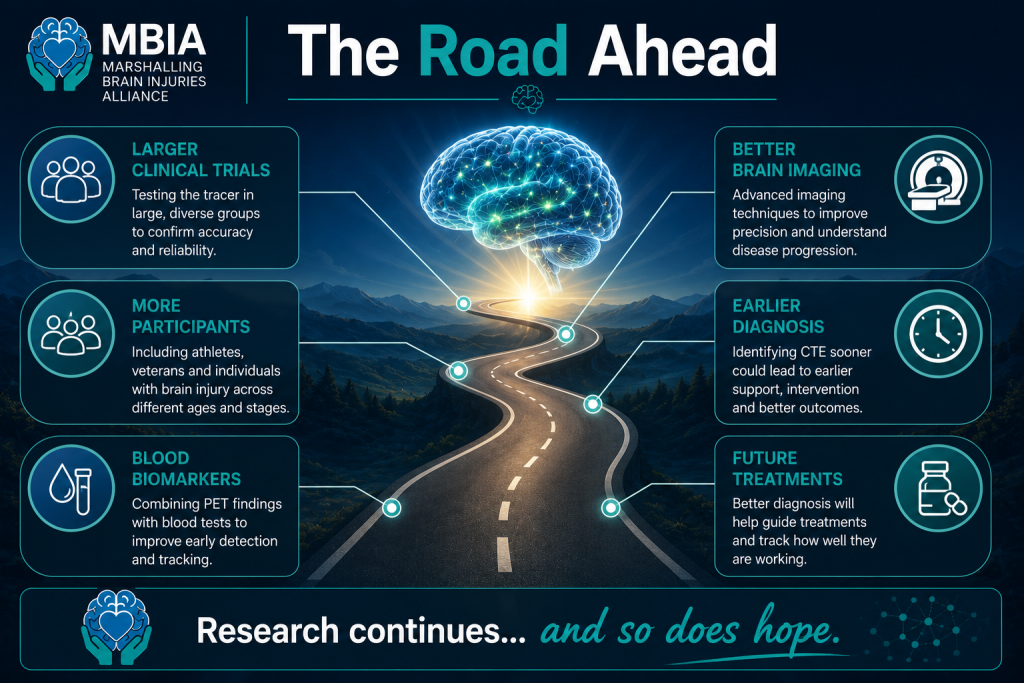
Researchers now need to carry out much larger clinical trials.
Future studies will likely include:
- More retired athletes
- Military veterans
- People with traumatic brain injury
- Comparison with Alzheimer's disease
- Long-term follow-up
- Brain imaging alongside blood biomarkers
Scientists are also working on combining PET imaging with blood tests, advanced MRI techniques and other biomarkers to improve the accuracy of diagnosis.
The future of CTE research is becoming increasingly collaborative.
MBIA's View
At Marshalling Brain Injuries Alliance, we believe this is one of the most promising developments in CTE research for several years.
It is far too early to describe this as a diagnostic test.
However, it demonstrates that researchers are making genuine progress towards identifying CTE during life.
That could transform not only research, but also how patients are diagnosed, supported and eventually treated.
We hope studies like this continue to receive funding and international collaboration.
Every breakthrough brings us one step closer to better answers for the thousands of individuals and families living with the consequences of repeated brain injury.
Key Takeaways
✔ Researchers tested a new PET scan tracer designed to detect CTE-related tau protein.
✔ The tracer successfully identified abnormal tau in a very small group of retired athletes and in donated CTE brain tissue.
✔ This could eventually help diagnose CTE during life, but much more research is needed.
✔ The study involved only three suspected CTE patients, so it is not yet ready for clinical use.
✔ Despite the limitations, this represents one of the most encouraging advances in CTE imaging research in recent years.

References
- Bieniek KF, et al. Early evaluation of the PET tracer 18F-OXD-2314 for detecting CTE-associated tau pathology. Presented at the Alzheimer's Association International Conference (AAIC), 2026.
- Alzheimer's Association International Conference (AAIC) 2026.
- EurekAlert! "New PET tracer shows promise for detecting CTE pathology in living people."
Enjoyed this article? Join thousands of readers receiving our monthly Brain Injury Brief