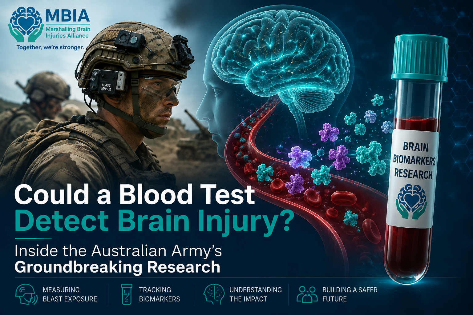
Could a Blood Test Detect Brain Injury? Inside the Australian Army's Ground breaking Research
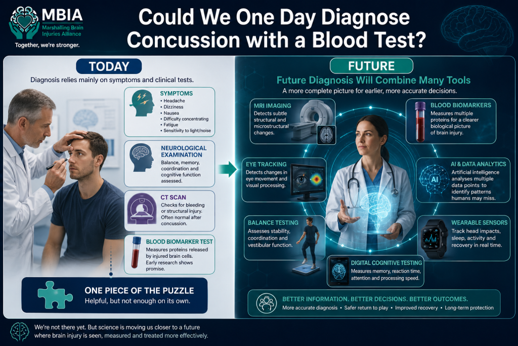
For decades, diagnosing a concussion has relied largely on something surprisingly subjective.
Doctors ask questions.
They assess symptoms.
They perform neurological examinations.
Sometimes a CT or MRI scan is performed—but these scans often appear completely normal after a mild traumatic brain injury.
This has left clinicians searching for something more objective.
What if a simple blood test could reveal whether the brain had been injured?
That question is now being investigated by researchers around the world, including the Australian Army, which has launched one of its largest studies into whether tiny proteins released into the bloodstream after brain injury could provide an earlier, more accurate picture of what's happening inside the brain.
If successful, the research could one day benefit not only soldiers exposed to blast waves, but also rugby players, footballers, emergency service workers and anyone exposed to repeated head impacts.
Why the Australian Army Is Interested
Before looking at the blood tests themselves, it's important to understand why the Australian Army is investing in this research.
When most people think about military brain injuries, they picture a large explosion causing an obvious traumatic brain injury. While these severe injuries remain a significant concern, researchers are becoming increasingly interested in something much less visible: the effects of repeated low-level blast exposure.
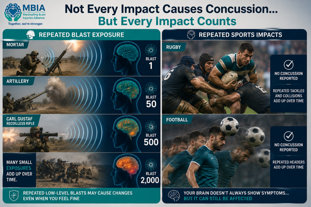
Many military personnel, particularly those who regularly train with heavy weapons such as artillery, mortars and recoilless rifles, are exposed to blast waves as part of their everyday duties. These blasts may not be powerful enough to knock someone unconscious or produce the classic signs of concussion. In fact, many soldiers continue training without experiencing any noticeable symptoms at all.
However, each blast generates a pressure wave that travels through the body and brain. Scientists are now asking whether repeated exposure to these pressure waves—sometimes occurring hundreds or even thousands of times over the course of a military career—could gradually affect brain health, even when no single event appears serious enough to be diagnosed as a concussion.
This idea is known as cumulative exposure. Rather than one major injury causing lasting damage, researchers are investigating whether many smaller exposures may add up over time, producing subtle changes that only become apparent months or years later.
This question has important similarities with contact sports.
In rugby, not every tackle causes a concussion. A player may make dozens of tackles during a single match and hundreds over the course of a season without ever being diagnosed with a head injury. Likewise, footballers may head the ball repeatedly without developing immediate symptoms, and boxers or mixed martial artists may absorb countless impacts that do not result in a recognised concussion.
Yet research over the past decade has suggested that the absence of symptoms does not necessarily mean the brain has been unaffected. Scientists are increasingly studying whether repetitive head impacts—including those that do not cause obvious concussion—may still trigger tiny biological changes within the brain.
This is why the Australian Army's research has attracted international attention. Instead of focusing solely on soldiers who have suffered diagnosed traumatic brain injuries, researchers are asking a much broader question:
Can repeated low-level blast exposure leave detectable biological signs of brain injury, even when a person feels completely well?
If blood biomarkers can identify these subtle changes, they could transform how military organisations monitor brain health. Importantly, the findings may also have implications far beyond the armed forces, helping researchers better understand the effects of repetitive head impacts in sports such as rugby, football and boxing, as well as in other occupations where repeated brain trauma is a concern.
Ultimately, the Australian Army's study reflects a growing shift in brain injury research. Instead of asking only "Did this person have a concussion?", scientists are beginning to ask a more complex—and potentially more important—question:
"What is the total effect of repeated impacts on the brain over an entire career?"
What Is a Brain Biomarker?
The word "biomarker" may sound technical, but the idea behind it is actually quite simple.
A biomarker is a measurable biological substance that provides information about what is happening inside the body. Doctors already use biomarkers every day. For example, blood glucose helps diagnose diabetes, cholesterol levels provide clues about heart disease risk, and troponin is released into the bloodstream when the heart muscle is damaged during a heart attack.
Researchers are now trying to identify similar biomarkers for the brain.
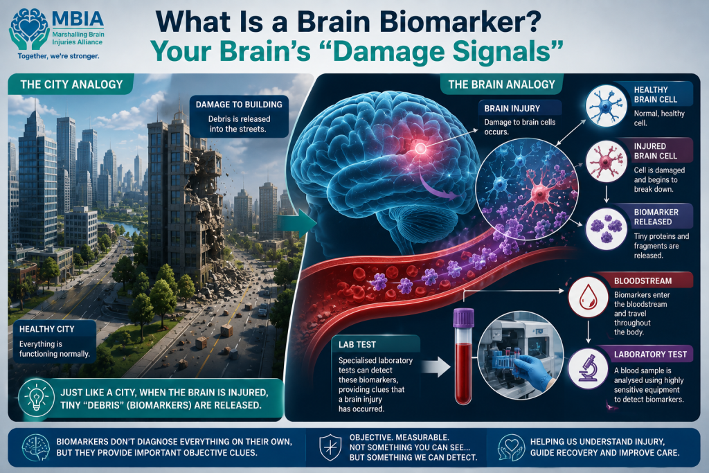
One way to understand this is to imagine your brain as a busy city.
The brain's billions of nerve cells are like buildings, roads and communication networks, all working together to keep the city running smoothly. Supporting cells act like maintenance crews, protecting and repairing the infrastructure while ensuring everything functions as it should.
If a building in the city is damaged, pieces of brick, glass and concrete spill out onto the surrounding streets. Even if you can't see the damaged building itself, the debris provides clear evidence that something has happened.
A similar process can occur inside the brain.
When brain cells or their supporting structures are injured—whether through a concussion, repeated head impacts or blast exposure—tiny proteins that are normally contained within those cells can escape into the surrounding fluid. Some of these proteins eventually cross into the bloodstream, where they can be detected using highly sensitive laboratory tests.
These proteins are known as brain biomarkers because they act as biological clues that an injury has occurred.
Importantly, the blood itself is not detecting the injury. Instead, scientists are measuring proteins that have been released by the injured brain. Think of them as microscopic "signals" that something inside the brain has changed.
This is what makes biomarkers so exciting.
Traditional concussion diagnosis relies heavily on symptoms such as headache, dizziness, confusion or memory problems. While these symptoms are important, they are also subjective. Some people may under-report symptoms because they want to keep playing sport or remain on duty. Others may struggle to describe exactly how they feel, particularly if the injury affects their thinking or communication.
Biomarkers are different.
They are objective biological measurements. A protein released into the bloodstream cannot exaggerate its level, hide itself, or forget what happened. It simply reflects the underlying biology occurring within the brain.
That doesn't mean biomarkers can tell doctors everything. A single blood test cannot currently diagnose every type of brain injury, determine how severe it is, or predict exactly how someone will recover. Researchers are still working to understand what different biomarker levels mean and how they change over time.
Nevertheless, these tiny proteins are offering scientists something they have long been searching for: an objective window into an injury that has often been invisible.
Rather than relying solely on what someone feels or what appears on a brain scan, biomarkers may eventually help clinicians detect, monitor and better understand brain injuries at a biological level.
How Can a Blood Test Detect Brain Injury?
At first glance, the idea of diagnosing a brain injury with a blood test seems almost impossible.
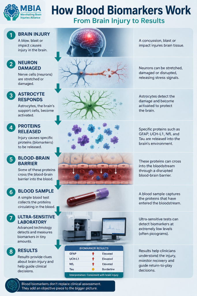
After all, the brain is protected inside the skull and separated from the rest of the body by a specialised defence system called the blood-brain barrier. This barrier acts like an extremely selective security checkpoint, controlling which substances can move between the bloodstream and the brain. Under normal circumstances, many proteins produced by brain cells remain almost entirely confined within the brain.
So how can a blood test reveal that the brain has been injured?
It begins with what happens at the moment of injury.
Whether the injury is caused by a fall, a concussion, repeated head impacts, or exposure to a blast wave, the brain's cells can experience physical stress. Nerve fibres (axons) may stretch, supporting cells can become damaged, and the brain's normal balance of chemicals is temporarily disrupted.
Almost immediately, the brain activates its natural defence and repair systems.
Supporting cells, including astrocytes and microglia, begin responding to the injury by helping clear away damaged tissue, regulating inflammation and attempting to restore normal brain function. At the same time, injured brain cells may release proteins that are normally kept safely inside them.
Some of these proteins gradually make their way into the fluid surrounding the brain. If the injury has disrupted the blood-brain barrier—even slightly—or enough protein has accumulated, small amounts can pass into the bloodstream.
These proteins are far too small and far too scarce to be detected by standard blood tests. In many cases, they are present at concentrations measured in trillionths of a gram per millilitre of blood.
This is where modern technology has transformed brain injury research.
Scientists now use highly sophisticated laboratory techniques, such as single molecule array (Simoa) technology and other ultra-sensitive assays, capable of detecting incredibly tiny amounts of these proteins with remarkable accuracy. Just a few decades ago, measuring concentrations this low would have been impossible.
It's important to understand what these tests are—and are not—doing.
The blood itself is not detecting the brain injury.
Instead, scientists are looking for proteins that have leaked from injured brain cells into the bloodstream. These proteins act like biological fingerprints, providing evidence that something has happened within the brain.
An analogy can help explain this.
Imagine a factory beside a river. If machinery inside the factory is damaged, traces of oil may eventually appear downstream. Testing the river doesn't reveal the damaged machine directly—it detects the substances that escaped because damage occurred.
Brain biomarker testing works in much the same way. Researchers aren't examining the brain itself; they're analysing the "downstream" evidence carried in the bloodstream.
The challenge is that no single protein tells the whole story. Different biomarkers reflect different types of brain cells and different aspects of injury. Some rise within minutes or hours, while others may remain elevated for days, weeks or even longer.
For this reason, researchers increasingly believe that the future lies not in a single "brain injury blood test," but in combining several biomarkers with clinical examination, cognitive testing and brain imaging to build a much more complete picture of what is happening inside the brain.
Although this science is still evolving, it represents one of the most promising advances in brain injury diagnosis for decades. For the first time, researchers can begin to measure the brain's response to injury through objective biological signals rather than relying solely on symptoms or scans that may appear completely normal.
Meet the Biomarkers
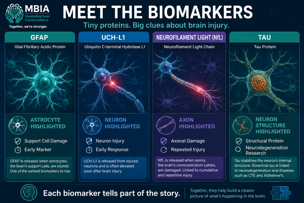
Scientists have identified dozens of proteins that may provide clues about brain injury, but a small number have emerged as the leading candidates for clinical use. Each biomarker tells researchers something slightly different about what is happening inside the brain. Rather than one protein providing all the answers, they are increasingly being viewed as pieces of a much larger puzzle.
GFAP (Glial Fibrillary Acidic Protein)
One of the most established brain injury biomarkers is Glial Fibrillary Acidic Protein, better known as GFAP.
GFAP is found primarily inside astrocytes, star-shaped support cells that play a vital role in maintaining the brain's health. Astrocytes help nourish neurons, regulate communication between brain cells, maintain the blood-brain barrier and respond rapidly when injury occurs.
When astrocytes are damaged, GFAP can leak into the bloodstream. One of the reasons researchers are particularly interested in GFAP is that it often rises relatively quickly after a brain injury, making it a useful marker during the early hours following trauma.
GFAP has already moved beyond the research laboratory. In the United States, it forms part of FDA-authorised blood tests used to help doctors decide whether someone with a mild traumatic brain injury is likely to have bleeding or other abnormalities visible on a CT scan. These tests can reduce unnecessary scans while helping identify patients who may require urgent imaging.
It's important to remember, however, that GFAP is not a concussion test and it cannot diagnose CTE. Instead, it provides one biological clue that brain cells supporting normal brain function may have been injured.
UCH-L1 (Ubiquitin C-Terminal Hydrolase L1)
Another important biomarker is UCH-L1, a protein found almost exclusively inside neurons, the brain's specialised nerve cells responsible for transmitting information throughout the nervous system.
When neurons are injured, UCH-L1 can escape into the bloodstream. Like GFAP, it often rises quite soon after an injury, making it particularly useful when assessing someone shortly after a suspected concussion or traumatic brain injury.
Because UCH-L1 reflects damage to neurons themselves rather than supporting cells, it provides scientists with a different perspective on what has happened inside the brain.
Researchers often measure GFAP and UCH-L1 together because the combination may offer a more complete picture of an injury than either biomarker alone.
Again, while UCH-L1 can indicate that neuronal injury has occurred, it cannot determine how severe the injury is, predict long-term recovery or diagnose conditions such as CTE by itself.
Neurofilament Light (NfL)
Among all the biomarkers currently being studied, Neurofilament Light, commonly shortened to NfL, has generated some of the greatest excitement.
Neurofilaments are structural proteins that act like the internal framework of nerve fibres, particularly the long extensions known as axons. These axons are responsible for carrying electrical signals between different parts of the brain and nervous system.
When axons are stretched or damaged—as can happen during concussion, repetitive head impacts or blast exposure—small amounts of NfL are released into the bloodstream.
Unlike some biomarkers that rise quickly and then fall within hours or days, NfL can remain elevated for much longer. This has made it particularly interesting for researchers studying whether repeated head impacts or repeated blast exposure may produce cumulative effects over time.
Studies involving rugby players, American football players, boxers, military personnel and people with traumatic brain injuries have all investigated NfL as a potential indicator of ongoing axonal damage.
Although the findings are promising, NfL is not specific to concussion. Levels can also increase in several neurological diseases, including multiple sclerosis, Alzheimer's disease, motor neurone disease and other conditions that damage nerve fibres. This means doctors must always interpret NfL results within the wider clinical picture.
Tau
Perhaps the best-known brain biomarker is tau, largely because it is closely associated with Alzheimer's disease and Chronic Traumatic Encephalopathy (CTE).
Under normal circumstances, tau performs an essential job inside neurons. It acts like scaffolding, helping stabilise the tiny structures that transport nutrients and other materials throughout the nerve cell.
Following brain injury, tau can become disrupted and released into the bloodstream. In some circumstances, abnormal forms of tau may accumulate inside the brain, where they can interfere with normal cell function.
Because abnormal tau deposits are one of the defining features seen in the brains of people with CTE after death, researchers have devoted enormous effort to studying whether blood tau levels might one day help identify people at risk while they are still alive.
However, the reality is more complex than many headlines suggest.
Tau levels can increase after concussion, but they can also change for many other reasons. Different forms of tau behave differently, and researchers are still trying to determine which are most useful. At present, no blood test measuring tau can diagnose CTE, nor can it reliably distinguish CTE from other neurological conditions.
Nevertheless, tau remains an important piece of the puzzle. When combined with other biomarkers such as GFAP, UCH-L1 and NfL, it may help researchers build a more detailed understanding of how different types of brain injury affect the brain over time.
Putting the Pieces Together
Each of these biomarkers reflects a different aspect of brain biology.
- GFAP tells us about injury to the brain's supporting astrocytes.
- UCH-L1 reflects damage to neurons.
- NfL provides insight into injury affecting axons, the brain's communication pathways.
- Tau helps researchers study changes linked to neuronal structure and neurodegeneration.
Rather than searching for one perfect biomarker, scientists increasingly believe that using several biomarkers together—alongside brain imaging, neurological examinations and cognitive testing—offers the greatest potential for improving the diagnosis and monitoring of brain injuries.
This "biomarker panel" approach is at the heart of many of today's most exciting studies, including the Australian Army's investigation into the effects of repeated blast exposure.
What Exactly Is the Australian Study Doing?
One of the reasons the Australian Army's research has attracted so much international attention is that it isn't simply taking blood samples from soldiers and hoping to find useful answers.
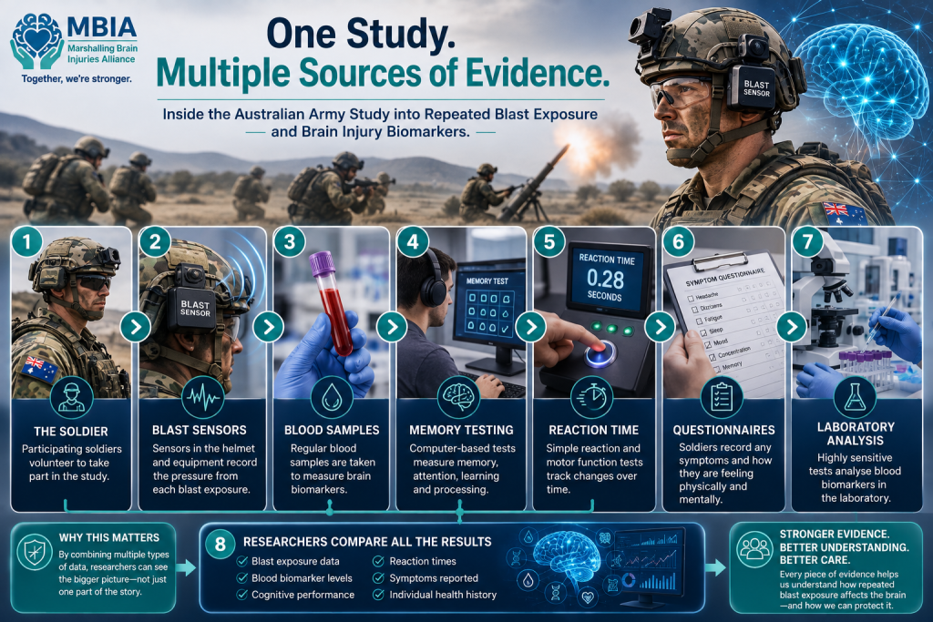
Instead, researchers are building a comprehensive picture of how repeated blast exposure affects the brain by combining physical measurements, biological testing, cognitive assessments and symptom reporting. Each piece of information helps answer a different part of the puzzle.
Measuring Blast Exposure
The first step is to understand exactly how much blast exposure each soldier experiences.
Participants wear specialised blast sensors attached to their helmets and equipment during training exercises involving heavy weapons such as artillery, mortars and recoilless rifles.
These sensors record the pressure generated by each blast wave, allowing researchers to measure not only whether a blast occurred, but also its intensity and how often soldiers are exposed over time.
Rather than relying on estimates or memory, scientists can collect objective data about an individual's cumulative blast exposure throughout the study.
Looking for Biological Changes
Alongside the physical measurements, researchers collect blood samples from participants.
These samples are analysed using ultra-sensitive laboratory techniques capable of detecting tiny concentrations of brain injury biomarkers such as GFAP, UCH-L1, Neurofilament Light (NfL) and tau.
The goal is to determine whether repeated blast exposure is followed by measurable changes in these proteins.
If certain biomarkers consistently increase after blast exposure, they may provide important biological evidence that the brain has responded to the pressure waves—even when no obvious concussion has occurred.
Testing Brain Function
The study isn't only interested in what happens in the blood.
Researchers also assess how well participants' brains are functioning through a range of cognitive tests designed to measure different aspects of thinking.
These assessments include tasks that evaluate:
- Memory
- Attention
- Concentration
- Processing speed
- Executive function
- Problem-solving ability
Many of these abilities can be subtly affected by brain injury, even when a person feels completely normal.
By repeating these assessments throughout the study, researchers can identify small changes that might otherwise go unnoticed in everyday life.
Measuring Reaction Time
Reaction time is another important part of the study.
The brain constantly receives information, processes it and sends instructions back to the body. Even very subtle disruption to these processes may slow how quickly someone responds.
Participants therefore complete reaction time tests to determine whether repeated blast exposure is associated with changes in the speed and efficiency of brain function.
While a fraction of a second may seem insignificant, consistent changes across a large group of participants can provide valuable scientific insights.
Listening to the Soldiers
Not every effect of brain injury can be measured by laboratory equipment.
For this reason, participants also complete detailed symptom questionnaires, reporting any headaches, dizziness, balance problems, fatigue, sleep disturbances, mood changes, concentration difficulties or memory concerns they experience during the study.
These self-reported symptoms help researchers understand how biological changes relate to real-world experiences.
A soldier may have no detectable symptoms despite measurable biomarker changes—or conversely, significant symptoms without major changes in a particular biomarker. Understanding these relationships is one of the study's key objectives.
Bringing Everything Together
Perhaps the most important aspect of the Australian Army's research is that no single test is being viewed in isolation.
Researchers will compare:
- The amount of blast exposure recorded by the helmet sensors.
- Changes in blood biomarker levels.
- Performance on memory and cognitive tests.
- Reaction times.
- Participants' reported symptoms.
By combining all of these data, scientists hope to determine whether repeated low-level blast exposure produces measurable biological changes, whether those changes affect thinking and memory, and whether blood biomarkers could eventually help identify soldiers at risk before more serious problems develop.
This comprehensive, multi-layered approach reflects the direction of modern brain injury research. Rather than searching for a single "magic test," scientists increasingly recognise that understanding the brain requires bringing together biology, technology, psychology and clinical assessment to create the clearest possible picture of what is happening beneath the surface.
If successful, this research could not only improve how military personnel are monitored but also provide valuable insights for athletes, emergency service workers and anyone exposed to repeated impacts to the head.
Why This Matters Beyond the Military
Although the Australian Army's study focuses on soldiers exposed to repeated blast waves, its significance extends far beyond the armed forces.
At its heart, the research is asking a much broader scientific question:
Can repeated forces acting on the brain leave behind measurable biological evidence, even when they don't cause an obvious concussion?
That question is relevant to millions of people around the world.
The source of the force may differ—a blast wave, a tackle, a punch or a fall—but the brain responds according to the same basic principles of biology. If brain cells are stretched, compressed or otherwise injured, they may release many of the same biomarkers into the bloodstream.
This is why researchers from so many different fields are watching studies like the Australian Army's with great interest.
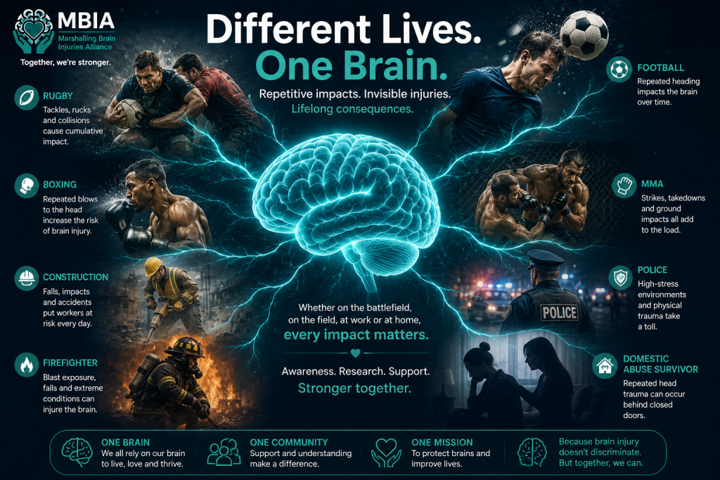
Rugby
For organisations like MBIA, the connection with rugby is particularly striking.
A rugby player may make dozens of tackles during a single match and many thousands throughout a career. Most of these impacts never result in a diagnosed concussion, and players often continue competing without any immediate symptoms.
However, researchers are increasingly investigating whether repetitive head impacts—even those below the threshold of concussion—may still cause subtle biological changes within the brain.
If blood biomarkers can detect these changes, they could one day help researchers better understand cumulative exposure and support safer decisions around player welfare, recovery and long-term brain health.
Football
Football presents similar questions.
Most headers do not cause concussion, and millions of players head the ball without experiencing immediate symptoms. Yet studies have raised concerns that years of repetitive heading may contribute to changes in brain structure and function for some players.
Blood biomarkers could provide researchers with a way of studying what is happening inside the brain after repeated heading, rather than relying solely on symptoms or brain scans.
Boxing and Mixed Martial Arts
Combat sports involve repeated blows to the head as an expected part of competition.
Even when an athlete is not knocked unconscious, punches and kicks can generate rapid acceleration and rotation of the brain inside the skull. Over time, these repeated impacts are believed to contribute to cumulative brain injury in some athletes.
Researchers are already studying biomarkers such as Neurofilament Light (NfL) and tau in professional boxers and mixed martial artists to better understand how repeated impacts affect brain health over months and years.
Construction and Other High-Risk Occupations
The military is not the only profession where workers may experience repeated physical forces affecting the head.
Construction workers, demolition crews, miners and others working with heavy machinery may be exposed to repeated vibration, accidental impacts or occasional blast exposure during their careers.
While these exposures differ from those experienced in combat, scientists are increasingly interested in whether long-term occupational exposure may also influence brain health.
Police and Firefighters
Police officers and firefighters routinely place themselves in dangerous environments.
Police officers may be assaulted, involved in high-speed collisions or exposed to repeated physical confrontations.
Firefighters can experience structural collapses, falling debris, explosions and other traumatic events during emergency responses.
Although brain injury research in these professions remains relatively limited, there is growing recognition that occupational head trauma deserves far greater attention.
Domestic Abuse Survivors
One of the most important applications of this research may be for survivors of domestic abuse.
Many survivors experience repeated blows to the head, strangulation or violent assaults over months or years. These injuries frequently go undocumented, and many victims never receive medical assessment at the time.
Research has increasingly shown that survivors of intimate partner violence may experience long-term neurological symptoms consistent with traumatic brain injury.
Objective biological markers could eventually help researchers better understand these injuries, improve recognition within healthcare settings and ensure survivors receive the support and rehabilitation they need.
Anyone Exposed to Repetitive Brain Trauma
Ultimately, the Australian Army's study is not just about soldiers.
It is about understanding how the human brain responds to repeated injury, regardless of where that injury occurs.
Whether someone is:
- a rugby player making hundreds of tackles,
- a footballer heading the ball throughout a career,
- a boxer stepping into the ring,
- a police officer responding to violent incidents,
- a firefighter attending dangerous emergencies,
- a construction worker exposed to repeated impacts,
- or a survivor of domestic abuse,
the underlying biology may have important similarities.
The exact forces acting on the brain may differ, but the cells responding to those forces are the same.
This is why advances in one area of brain injury research often benefit many others.
The Australian Army's work may be focused on blast exposure today, but the knowledge gained could help improve concussion research in sport, enhance care for survivors of domestic abuse, inform occupational health policies and deepen our understanding of repetitive brain trauma across society.
Rather than treating military injuries, sports injuries and civilian brain injuries as completely separate problems, researchers are increasingly recognising that they are connected by the same underlying biological processes.
Understanding those shared processes may ultimately lead to better diagnosis, more personalised treatment and improved protection for everyone at risk of brain injury—not just those serving on the battlefield.
One of the biggest questions people ask when they hear about brain biomarkers is:
"Does this mean we'll soon have a blood test that can diagnose concussion?"
The honest answer is not yet—but we're getting closer.
Despite some sensational headlines, there is currently no blood test that can reliably diagnose every concussion, determine how severe it is, or predict exactly how someone will recover. Concussion remains a clinical diagnosis, meaning doctors still rely on a person's symptoms, neurological examination, medical history and, when appropriate, brain imaging to make decisions about diagnosis and treatment.
However, that doesn't mean blood biomarkers aren't already making a difference.
Blood Biomarkers Are Already Being Used Clinically
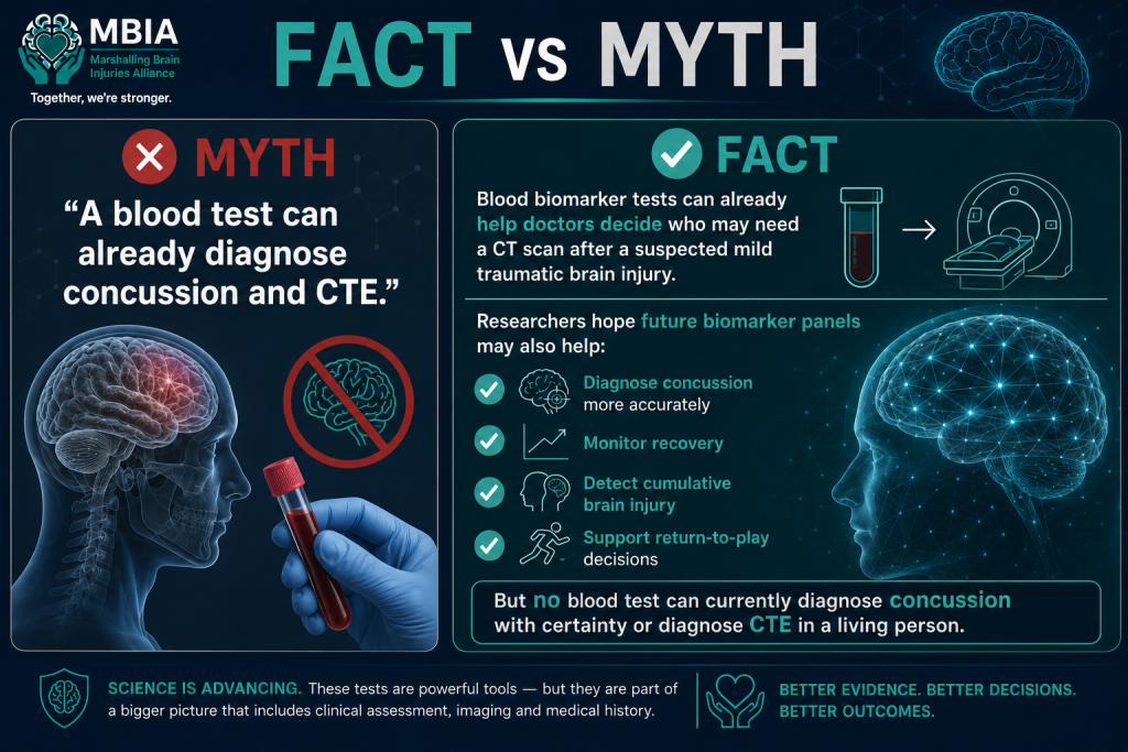
In the United States, the U.S. Food and Drug Administration (FDA) has authorised blood biomarker tests that measure proteins such as GFAP and UCH-L1 in people with a suspected mild traumatic brain injury.
These tests are not designed to diagnose concussion itself.
Instead, they help doctors estimate whether a patient is likely to have bleeding or other abnormalities that would be visible on a CT scan. If the biomarker levels are very low, the likelihood of finding a significant brain injury on a CT scan is also low, meaning some patients may be able to avoid unnecessary exposure to radiation and lengthy hospital investigations.
This represents an important advance, but it is only one step along the journey.
Where Researchers Hope the Science Will Go
Scientists believe the real potential lies in developing biomarker panels—combinations of several different proteins measured together rather than relying on a single marker.
Because each biomarker reflects a different aspect of brain injury, combining them could provide a much more complete picture of what is happening inside the brain.
Researchers hope that future biomarker panels may eventually help clinicians:
- Diagnose concussion more accurately, particularly when symptoms are unclear or delayed.
- Monitor recovery, allowing doctors to see whether the brain is healing alongside improvements in symptoms.
- Identify cumulative brain injury caused by repeated head impacts or blast exposure, even when no single event results in a diagnosed concussion.
- Support return-to-play or return-to-duty decisions, alongside clinical assessment, cognitive testing and other investigations.
Rather than replacing doctors, these tests could become another valuable tool to support clinical decision-making.
Why It's So Challenging
Developing a true concussion blood test is far more complicated than many people realise.
No two brain injuries are exactly alike. The amount of force involved, the area of the brain affected, a person's age, medical history, previous brain injuries and even the timing of the blood sample can all influence biomarker levels.
Some proteins rise within minutes before falling rapidly. Others increase more slowly and may remain elevated for days or weeks. Certain biomarkers can also rise because of neurological diseases unrelated to concussion, making interpretation more difficult.
This is why researchers increasingly believe that there is unlikely to be one single "magic" biomarker that provides all the answers.
Instead, the future probably lies in combining multiple biomarkers with brain imaging, cognitive testing, balance assessments, eye movement analysis and detailed clinical examination to build the clearest possible picture of an individual's brain health.
The Current Reality
The excitement surrounding blood biomarkers is entirely justified—they represent one of the most promising advances in brain injury research for decades.
But it's equally important to separate hope from hype.
At present:
- There is no blood test that can definitively diagnose concussion.
- There is no blood test that can determine how severe a concussion is.
- There is no blood test that can predict exactly how long recovery will take.
- There is no blood test that can diagnose Chronic Traumatic Encephalopathy (CTE) in a living person.
The Australian Army's study, along with many others around the world, is helping researchers move closer to these goals. Every new study improves our understanding of how the brain responds to injury and how biomarkers behave over time.
While we are not yet at the point where a simple blood test can answer every question about concussion, the science is advancing rapidly. The hope is that, in the future, blood biomarkers will become part of a comprehensive assessment—helping clinicians make more informed decisions and ensuring that people with brain injuries receive faster, more accurate and more personalised care.
The Bigger Picture
Although blood biomarkers are one of the most exciting developments in brain injury research, few scientists believe they will ever provide all the answers on their own.
The brain is the most complex organ in the human body. A single blood test can reveal part of the story, but it cannot show exactly where an injury has occurred, how it is affecting brain function, or how it may change over time.
Instead, researchers increasingly believe the future of brain injury diagnosis will involve bringing together multiple sources of information, each contributing a different piece of the puzzle.
Rather than asking one test to do everything, clinicians may one day combine biological, structural, functional and behavioural data to build a far more complete picture of an individual's brain health.
MRI: Looking at Brain Structure
Magnetic Resonance Imaging (MRI) has been used for decades to examine the brain's anatomy.
Conventional MRI is excellent for detecting problems such as bleeding, tumours or large areas of damage. However, many people with concussion or mild traumatic brain injury have scans that appear completely normal.
Researchers are therefore developing more advanced MRI techniques, such as Diffusion Tensor Imaging (DTI) and Magnetic Resonance Elastography (MRE), which may be able to detect subtle changes in white matter pathways and the brain's mechanical properties that standard MRI cannot reveal.
Future blood biomarker results could be interpreted alongside these advanced imaging techniques to provide both biological and structural evidence of injury.
PET Scans: Looking at Brain Biology
While MRI shows what the brain looks like, Positron Emission Tomography (PET) helps researchers study how the brain is functioning at a molecular level.
Special radioactive tracers can be used to investigate processes such as inflammation, glucose metabolism and, increasingly, the abnormal accumulation of proteins such as tau.
PET imaging is already playing an important role in Alzheimer's disease research and is also being investigated as a potential tool for identifying CTE-related changes during life.
Although PET scans remain primarily a research tool for repetitive brain injury, combining PET findings with blood biomarkers may eventually provide a more detailed understanding of ongoing disease processes.
Eye Movement Testing
Our eyes provide a remarkable window into brain function.
The brain controls eye movements using networks spread across multiple regions. Even mild brain injuries can affect how smoothly the eyes track moving objects, coordinate together or respond to visual information.
Modern eye-tracking technology can detect tiny abnormalities that may not be obvious during a routine examination.
Researchers are exploring whether combining eye movement testing with blood biomarkers could improve the early detection and monitoring of concussion.
Balance Testing
Maintaining balance requires constant communication between the brain, inner ear, eyes and muscles.
Following a concussion, these systems may not work together as efficiently, leading to dizziness, unsteadiness or poor coordination.
Computerised balance assessments can measure these changes far more accurately than simple bedside tests.
When interpreted alongside blood biomarkers, balance testing may provide additional evidence of how a brain injury is affecting day-to-day function.
Speech and Voice Analysis
One of the newest areas of research involves analysing the way people speak.
Changes in speech rate, pauses, word choice, pronunciation and vocal characteristics may reflect subtle alterations in brain function that are difficult for clinicians to detect during a normal conversation.
Artificial intelligence can already identify patterns in speech that humans might miss, and researchers are investigating whether these changes could help detect neurological disorders, monitor recovery or identify early signs of cognitive decline.
Although still in its infancy, speech analysis could become another valuable component of future brain health assessments.
Digital Cognitive Testing
Traditional pen-and-paper memory tests are increasingly being replaced or supplemented by digital assessments.
Computer-based cognitive tests can measure reaction time, attention, memory, processing speed and executive function with remarkable precision. They also allow repeated testing over time, making it easier to monitor changes during recovery.
Rather than relying solely on how someone feels, clinicians may eventually compare symptom reports with objective changes in cognitive performance and blood biomarker levels.
Wearable Sensors
Technology is also transforming how head impacts are measured.
Modern helmet sensors, instrumented mouthguards and wearable devices can record the number, location and force of impacts experienced during sport or military training.
Instead of estimating exposure based on memory, researchers can collect accurate data about every significant impact a person experiences.
This information can then be compared with changes in blood biomarkers, cognitive performance and symptoms to better understand how repeated impacts affect the brain over time.
Artificial Intelligence
Perhaps the most powerful tool of all will be artificial intelligence (AI).
The challenge facing clinicians is not a lack of information—it's making sense of enormous amounts of complex data.
An individual may have blood biomarker results, MRI scans, PET imaging, eye movement data, balance assessments, speech analysis, cognitive test scores, wearable sensor data and detailed clinical notes. No human can easily identify every meaningful relationship between all of these variables.
AI systems are increasingly being developed to analyse these large datasets, identify hidden patterns and help clinicians make more accurate predictions about diagnosis, recovery and long-term outcomes.
Importantly, AI is unlikely to replace healthcare professionals. Instead, it has the potential to become a powerful decision-support tool, helping doctors interpret information that would otherwise be impossible to analyse in full.
The Future Is Integration, Not Isolation
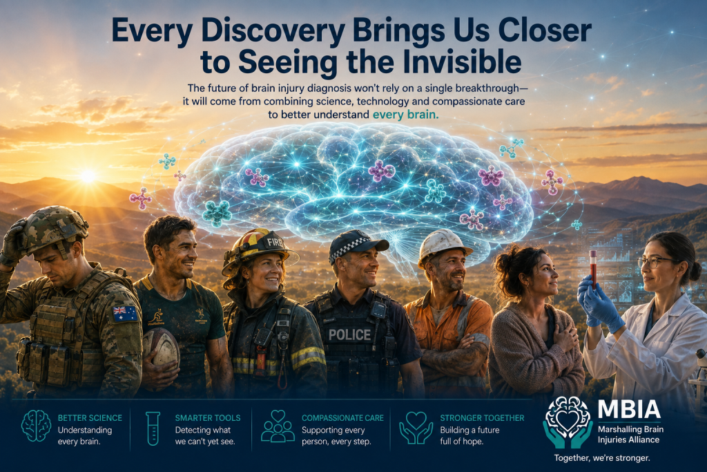
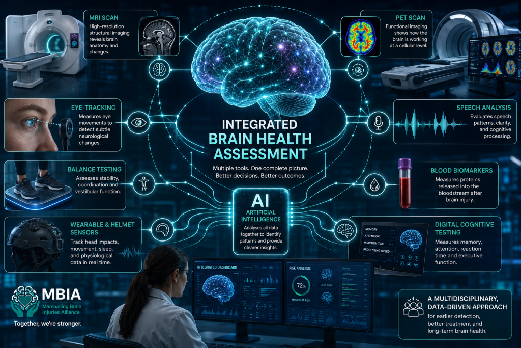
The future of brain injury diagnosis is unlikely to depend on one revolutionary test.
Instead, it will almost certainly involve integrating many different technologies, each providing a unique perspective on brain health.
Blood biomarkers may reveal the brain's biological response to injury.
MRI and PET scans may show structural and molecular changes.
Eye movement, balance and speech analysis may detect subtle functional impairments.
Wearable sensors may quantify the forces experienced by the brain.
Digital cognitive tests may measure how those changes affect thinking and memory.
Artificial intelligence may bring all of these pieces together into a single, personalised assessment.
No single technology will provide every answer. But together, they may allow clinicians to see brain injuries with a level of accuracy that would have seemed impossible just a decade ago.
For people living with brain injury, that future offers genuine hope—not because one test will solve every problem, but because combining multiple tools could lead to earlier diagnosis, more personalised treatment and better-informed decisions throughout recovery.