What Is Photobiomodulation? Could Light Therapy Help the Brain?
Introduction
Many people living with brain injuries, persistent concussion symptoms or dementia have heard claims that red light therapy or photobiomodulation (PBM) can "repair the brain."
Some describe it as revolutionary.
Others dismiss it as pseudoscience.
So what does the research actually say?
The answer is more interesting—and far more nuanced—than many headlines suggest.
Scientists have discovered that specific wavelengths of red and near-infrared light can trigger biological changes inside cells. Rather than heating tissue, this light appears to influence how cells produce energy, respond to inflammation and repair themselves after injury.
Researchers are now investigating whether these effects could benefit people living with traumatic brain injury (TBI), persistent post-concussion symptoms, Alzheimer's disease and other neurological conditions.
The early findings are encouraging—but many important questions remain unanswered.
In this article we separate the science from the hype and explain what photobiomodulation is, how it works and what the evidence really tells us today.
Light therapy that supports the brain's natural biology—not science fiction.
When most people first hear the word photobiomodulation (pronounced foe-toe-bio-mod-you-lay-shun), it sounds like something from a futuristic film.
Light treating the brain?
How could that possibly work?
The reality is both much simpler—and much more scientifically interesting.
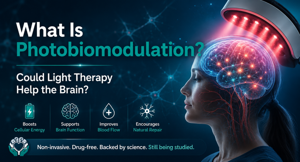
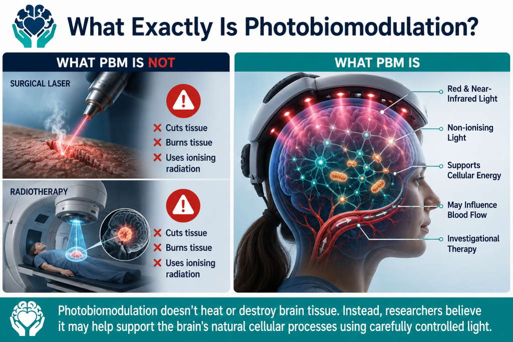
Photobiomodulation, usually shortened to PBM, is a form of light therapy that uses carefully controlled red and near-infrared light to influence how cells function. Unlike the powerful lasers used in surgery, PBM does not cut, burn or heat tissue. Instead, it delivers specific wavelengths of light at relatively low power, allowing cells to absorb the light and trigger a series of natural biological responses.
Scientists sometimes describe this as encouraging the body's own repair and maintenance systems rather than forcing them to do something unnatural.
Think of it like watering a plant.
The water doesn't make the plant grow directly. Instead, it provides the conditions the plant needs to grow itself.
Photobiomodulation works in a similar way. The light isn't "fixing" damaged brain cells. Instead, researchers believe it may help cells function more efficiently by supporting the biological processes they already use every second of every day.
Why Isn't It Like Laser Surgery?
Many people hear the word laser and immediately think of operations where surgeons cut through tissue using intense beams of light.
Photobiomodulation is completely different.
Surgical lasers use extremely high-powered light that generates enough heat to cut, burn or remove tissue with great precision. They are designed to destroy tissue in a controlled way.
Photobiomodulation uses low-intensity light that produces little or no heating of the tissue. Rather than damaging cells, the goal is to influence their chemistry.
You can think of the difference like sunlight and a magnifying glass.
A magnifying glass concentrates sunlight enough to burn paper.
Ordinary sunlight, however, simply provides light and warmth without causing damage.
PBM uses light much more like the second example—it acts as a biological signal rather than a cutting tool.
Is It Like Radiotherapy?
No.
This is another common misunderstanding.
Radiotherapy uses ionising radiation, such as high-energy X-rays or gamma rays, to damage the DNA inside cancer cells so they can no longer grow.
Photobiomodulation uses non-ionising light, usually in the red or near-infrared part of the light spectrum.
Non-ionising light does not carry enough energy to damage DNA in the way ionising radiation does. Instead, it interacts with light-sensitive molecules already present inside our cells.
In other words, radiotherapy aims to destroy harmful cells.
Photobiomodulation aims to support the normal function of healthy or stressed cells.
These are fundamentally different treatments that simply happen to involve light.
LEDs and Lasers – What's the Difference?
One of the biggest myths surrounding photobiomodulation is that every treatment uses lasers.
In reality, modern PBM devices may use either lasers or light-emitting diodes (LEDs).
Both can produce the specific wavelengths of red or near-infrared light used in research.
The main difference is how that light is delivered.
Lasers produce highly focused, coherent beams of light that travel in a single direction. They can deliver light very precisely to a small area.
LEDs, on the other hand, produce light that spreads over a wider area. This makes them particularly useful for treating larger parts of the body, such as the scalp.
An easy way to picture the difference is to imagine two torches.
A laser is like a narrow spotlight shining on a single object.
An LED panel is more like a floodlight that gently illuminates a much larger area.
Importantly, researchers have found that both technologies can produce biological effects when the correct wavelengths and treatment parameters are used. This is why many modern brain PBM systems use arrays of LEDs rather than lasers alone.
Why Do Scientists Use Near-Infrared Light?
Not all light travels through the body equally well.
Visible red light can penetrate the skin and some of the tissues beneath it, which is why it is widely used for skin health and wound healing.
Near-infrared light, however, has a special advantage.
Although it is invisible to the human eye, it can travel further through skin, connective tissue and bone before being absorbed.
That makes it more suitable for researchers investigating treatments aimed at the brain.
Imagine shining a torch through your hand.
Some colours of light are quickly blocked.
Others pass much further through the tissue before fading away.
Near-infrared light behaves a little like the second example—it can penetrate deeper than visible red light, although only a small proportion of the original light reaches the brain itself. The exact amount varies depending on many factors, including the wavelength used, the power of the device, skull thickness, hair, skin and the area being treated.
This is an important point.
Some advertisements give the impression that near-infrared light easily reaches every part of the brain.
That isn't what the research shows.
Scientists agree that much of the light is absorbed by the scalp and skull before it reaches brain tissue. The question researchers are now trying to answer is whether the small amount that does reach the brain is enough to trigger meaningful biological effects.
So far, early studies suggest it may be—but this remains one of the biggest areas of ongoing research.
Understanding why the brain's enormous energy demands make it vulnerable to injury and disease.
When people think about the body's biggest energy users, they often imagine the heart constantly pumping blood or the powerful muscles in our legs carrying us through the day.
Surprisingly, one of the body's most energy-hungry organs is actually the brain.
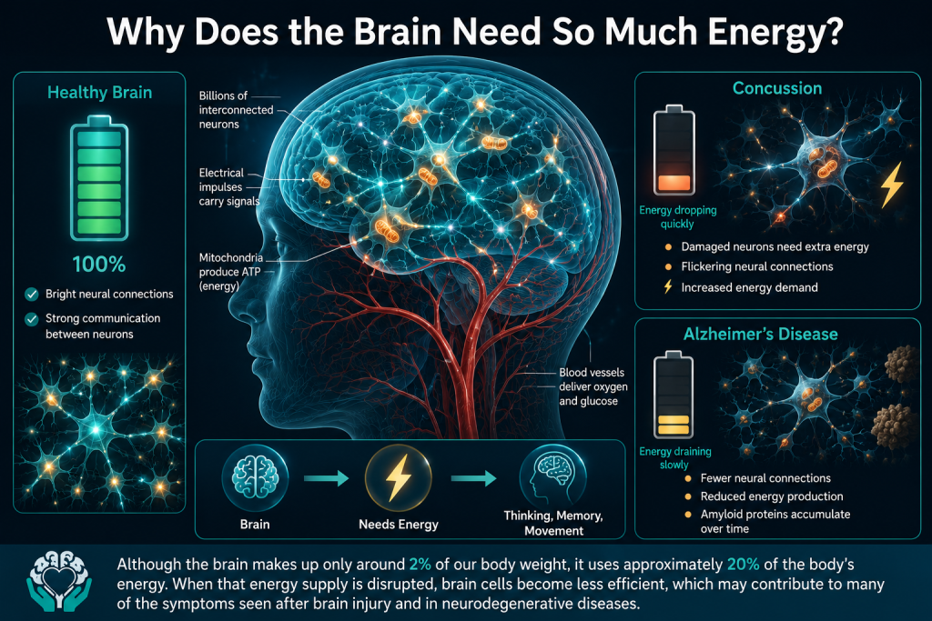
Although the brain accounts for only around 2% of our body weight, it consumes approximately 20% of the body's total energy and oxygen supply while we are at rest. In other words, gram for gram, your brain uses far more energy than almost any other organ in your body.
That energy is needed every second of every day.
Unlike a computer that can simply be switched off when it's not being used, the brain is constantly active. Even while you're asleep, billions of nerve cells are communicating, repairing themselves, storing memories, regulating your heartbeat, controlling your breathing and processing information from the world around you.
It's an incredibly demanding job.
Your Brain Is Like a City That Never Sleeps
One way to understand the brain's energy needs is to imagine a large city.
Every house needs electricity.
Traffic lights have to keep working.
Hospitals cannot lose power.
Mobile phone networks must stay connected.
Water has to be pumped continuously.
Now imagine if the city's power stations suddenly began producing less electricity.
At first, the lights might flicker.
Traffic would slow.
Computers would struggle to work properly.
Communication systems would become unreliable.
Eventually, if the shortage continued, parts of the city would begin shutting down altogether.
The brain behaves in a remarkably similar way.
When brain cells don't have enough energy, they don't immediately die. Instead, they often become less efficient, making it harder for them to communicate with one another. This can affect memory, concentration, mood, decision-making and the ability to process information—symptoms that many people living with brain injuries or neurodegenerative diseases know all too well.
Neurons: Tiny Electrical Factories Working Around the Clock
The brain contains an estimated 86 billion neurons, each connected to thousands of other nerve cells through tiny junctions called synapses.
Every thought you have...
Every word you speak...
Every memory you recall...
Every movement you make...
depends on these neurons sending tiny electrical and chemical signals across incredibly complex networks.
But producing these signals isn't free.
Each neuron is like a miniature electrical factory, continuously using energy to:
- Generate electrical impulses.
- Send chemical messages to neighbouring cells.
- Repair and maintain its internal structures.
- Remove waste products.
- Build and strengthen new connections during learning.
- Respond to injury or illness.
Unlike many other cells in the body, neurons have very little ability to "rest." They are active almost constantly, meaning they need a reliable supply of energy every second.
Even a short interruption in energy production can begin affecting how well they function.
What Happens After a Brain Injury?
One of the first things that happens after a concussion or traumatic brain injury (TBI) is something scientists often call an energy crisis.
When the brain is exposed to a sudden impact or rapid acceleration and deceleration, millions of neurons are stretched and disrupted.
To restore normal function, these injured cells suddenly need far more energy than usual.
At exactly the same time, however, the systems responsible for producing that energy become less efficient.
It's a bit like trying to recharge your phone while the charger itself has been damaged.
The phone desperately needs power, but the charging system isn't working properly.
Researchers have found that after concussion, brain cells can experience:
- Reduced energy production.
- Increased demand for fuel.
- Disturbances in calcium levels inside cells.
- Increased oxidative stress.
- Inflammation.
- Reduced blood flow in some parts of the brain.
This mismatch between energy supply and energy demand may help explain why many people experience symptoms such as:
- Mental fatigue.
- Brain fog.
- Poor concentration.
- Slower thinking.
- Headaches.
- Sensitivity to light and noise.
For some people these changes resolve within days or weeks.
For others, particularly after repeated injuries or more severe trauma, the brain may struggle to fully restore its normal energy balance.
Energy Problems Are Also Seen in Alzheimer's Disease
Scientists have discovered that similar energy problems occur in Alzheimer's disease, although they develop much more gradually.
Years before significant memory loss becomes obvious, researchers have observed that certain parts of the brain begin using less glucose—the brain's main source of fuel.
Brain imaging studies have also identified changes in:
- Mitochondrial function.
- Oxygen use.
- Oxidative stress.
- The way neurons produce and use energy.
Exactly what comes first is still debated.
Some researchers believe these energy problems may help drive the disease.
Others think they occur because of the build-up of amyloid proteins, tau tangles and inflammation.
Many now believe the relationship is likely to work both ways, with each problem making the others worse over time. Rather than being caused by a single factor, Alzheimer's disease appears to involve a complex cycle of interconnected biological changes.
Why Are Scientists So Interested in Cellular Energy?
For decades, much of Alzheimer's research focused on removing amyloid plaques from the brain.
While this remains an important area of research, scientists have increasingly recognised that keeping brain cells healthy may be just as important as removing harmful proteins.
If researchers can find safe ways to help neurons produce energy more efficiently, improve blood flow and reduce cellular stress, they may be able to support brain function even before significant damage occurs.
This is one reason photobiomodulation has attracted so much scientific interest.
Rather than targeting a single protein, researchers believe it may influence several of the brain's natural repair and maintenance systems at the same time.
Whether those biological changes lead to meaningful improvements for people living with brain injuries or dementia is the question that scientists are now working hard to answer.
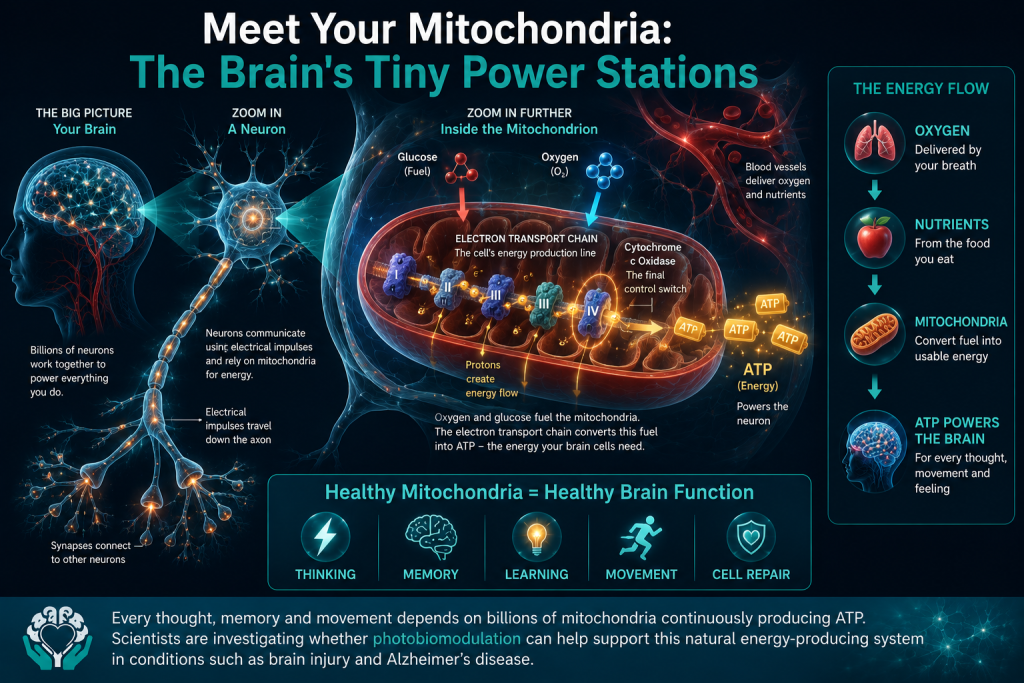
How billions of microscopic structures keep every thought, memory and movement alive.
Now that we understand just how much energy the brain needs, the next question is obvious.
Where does all that energy come from?
The answer lies inside almost every cell in your body.
Tiny structures called mitochondria (pronounced my-toe-CON-dree-ah).
Although they're far too small to see without a powerful microscope, mitochondria are some of the hardest-working parts of the human body. Every neuron in your brain contains hundreds—sometimes even thousands—of them, all working around the clock to generate the energy needed to keep your brain functioning.
Without healthy mitochondria, brain cells simply cannot do their job.
This is one reason why scientists have become so interested in them. Research has shown that mitochondrial function can become disrupted after traumatic brain injury, concussion and in neurodegenerative diseases such as Alzheimer's disease. If these tiny "power stations" begin struggling, the brain may gradually lose its ability to produce the energy it needs to think, learn, remember and repair itself.
What Are Mitochondria?
Imagine every cell in your brain is a busy factory.
Inside each factory are tiny power stations supplying electricity to every machine.
Those power stations are the mitochondria.
Their job is remarkably simple—but incredibly important.
They take the oxygen you breathe and the nutrients from the food you eat, then convert them into a form of usable energy that your cells can actually use.
Without them, every biological process inside the cell would grind to a halt.
This is why mitochondria are often called the powerhouses of the cell.
It's a nickname that's stuck for decades because it captures their essential role so well.
But mitochondria do much more than simply produce energy.
Scientists now know they also help regulate inflammation, control calcium levels inside cells, produce important signalling molecules, defend against oxidative stress and even help determine whether damaged cells survive or die.
In other words, they're not just batteries.
They're more like intelligent energy management centres, constantly monitoring what the cell needs and responding to changing conditions.
ATP – The Brain's Energy Currency
The energy produced by mitochondria isn't stored as electricity.
Instead, it's packaged into a tiny molecule called ATP, short for adenosine triphosphate.
You don't need to remember the name.
What's important is understanding what ATP does.
Think of ATP as the brain's version of money.
Just as every purchase in a shop requires money, every task performed by a brain cell requires ATP.
Every electrical signal travelling between neurons...
Every memory being stored...
Every muscle movement...
Every repair process...
Every new brain connection formed during learning...
All of these activities "cost" ATP.
Your brain is constantly spending this energy currency—and your mitochondria are constantly making more.
In fact, your body recycles its entire supply of ATP thousands of times every day.
That's why even a small disruption in energy production can quickly affect how well brain cells work.
How Do Mitochondria Make ATP?
This is where things become fascinating.
Inside every mitochondrion is an incredibly efficient microscopic production line known as the electron transport chain.
Despite the complicated name, the basic idea is surprisingly easy to understand.
Imagine a hydroelectric power station.
Water flows downhill through a series of turbines.
As the water moves, each turbine helps generate electricity.
The electron transport chain works in a similar way.
Instead of water, it uses tiny charged particles called electrons.
These electrons are passed from one protein to another in a carefully organised chain—rather like runners passing a relay baton.
Each handover releases a small amount of energy.
Rather than letting that energy go to waste, the mitochondrion captures it and uses it to build ATP.
By the time the process finishes, oxygen combines with the remaining electrons to form water—a perfectly normal by-product of cellular respiration.
This remarkable system is happening inside billions of cells throughout your body every second of every day.
Without it, life simply wouldn't be possible.
Meet Cytochrome c Oxidase – The Enzyme at the Centre of the Story
One of the final—and most important—proteins in this energy production line is an enzyme called cytochrome c oxidase.
Don't worry about remembering the name.
Think of it as one of the main control switches inside the mitochondrion.
Its job is to help the final stages of ATP production run smoothly by allowing oxygen to be used efficiently.
This is the protein that has attracted so much interest in photobiomodulation research.
Scientists believe that red and near-infrared light may be absorbed by cytochrome c oxidase, allowing it to work more efficiently under certain conditions. This could potentially improve how mitochondria produce ATP and may also influence blood flow, inflammation and other important cellular signalling pathways.
However, it's important to be clear about what the evidence shows.
Many laboratory studies support this theory, and cytochrome c oxidase remains the leading proposed target for photobiomodulation.
But researchers are still debating whether it explains all of the biological effects seen with red and near-infrared light.
Other light-sensitive molecules and cellular pathways may also play important roles.
In science, it's perfectly normal for several mechanisms to contribute to the same effect—and this is exactly what many researchers now suspect is happening with photobiomodulation.
Why Does This Matter After Brain Injury?
Following a concussion or traumatic brain injury, brain cells suddenly need enormous amounts of ATP to repair damaged membranes, restore normal electrical activity and recover from the injury.
Unfortunately, this often happens at exactly the same time that mitochondrial function has been disrupted.
Imagine a city hit by a major storm.
Thousands of engineers are urgently needed to repair damaged roads and restore electricity.
But many of the city's power stations have also been damaged.
The repairs become much harder because there simply isn't enough power available.
Researchers believe something similar may happen inside injured brain cells.
The demand for energy increases dramatically just as the cell's ability to produce ATP becomes less efficient.
This "energy mismatch" is thought to contribute to many of the symptoms experienced after brain injury, including fatigue, slower thinking and difficulties with memory and concentration.
Because photobiomodulation appears to target mitochondrial function, scientists are investigating whether supporting these tiny power stations could help brain cells recover more effectively.
Early laboratory and human studies have produced encouraging results, but researchers are still working to determine exactly how much benefit this may provide in real-world clinical practice.
Why Scientists Are Excited About Mitochondria
For many years, brain injury and Alzheimer's research focused primarily on the damage that occurs after disease or trauma.
Today, scientists are increasingly asking a different question.
Can we help brain cells function better before they become permanently damaged?
If therapies such as photobiomodulation can safely improve how mitochondria produce energy—or support the brain's natural repair processes—they could represent an entirely different approach to treating neurological disease.
Instead of targeting a single protein or symptom, they may help strengthen the brain's own ability to cope with stress and injury.
That's an exciting possibility.
But it's still exactly that—a possibility.
The challenge now is proving through large, carefully designed clinical trials whether improving mitochondrial function translates into meaningful improvements in memory, thinking, recovery and quality of life for people living with brain injuries and neurodegenerative diseases.
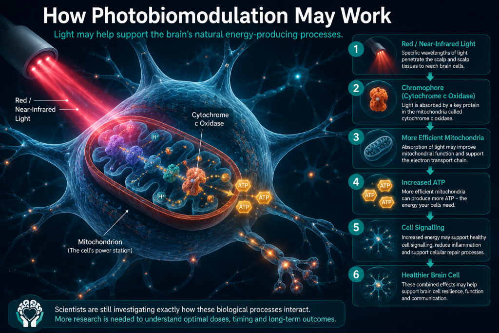
How Can Light Affect Cells?
The fascinating science behind photobiomodulation—and why researchers are still working to understand exactly how it works.
By now, we've seen that the brain relies on an enormous amount of energy and that tiny structures called mitochondria are responsible for producing much of it.
So where does light fit into the picture?
This is one of the most exciting—and most carefully debated—areas of photobiomodulation research.
Scientists agree that red and near-infrared light can produce measurable biological effects inside cells. Laboratory experiments and human studies have shown changes in cellular activity, blood flow, oxygen use and brain function after exposure to carefully controlled light.
What researchers are still trying to determine is exactly how these changes happen.
The leading theory involves mitochondria, but it is unlikely to be the whole story.
Rather than thinking of photobiomodulation as flipping a single switch, many scientists now believe it acts more like an orchestra conductor—gently influencing several biological systems that work together to help cells function more effectively.
Step 1: Light Meets a Cellular "Light Sensor"
Every colour of light carries a different amount of energy.
Some wavelengths pass straight through our tissues.
Others are absorbed by specific molecules inside our cells.
These light-absorbing molecules are known as chromophores.
You can think of a chromophore as a tiny solar panel.
Just as solar panels capture sunlight and convert it into electricity, chromophores capture certain wavelengths of red or near-infrared light and convert them into biological signals.
Researchers believe one of the most important chromophores inside our cells is an enzyme called cytochrome c oxidase, found within mitochondria.
When this enzyme absorbs light, it may begin a chain of events that influences how the cell produces energy and responds to stress.
However, scientists are careful not to describe this as established fact.
Although cytochrome c oxidase remains the leading proposed target, several recent studies suggest other light-sensitive molecules may also contribute to the effects of photobiomodulation. Rather than there being one single "light switch," multiple biological pathways may be involved simultaneously.
Step 2: Helping the Cell Produce Energy
If cytochrome c oxidase becomes more active, researchers believe the mitochondria may become temporarily more efficient at producing ATP—the energy molecule that powers every brain cell.
Remember our earlier analogy.
The light isn't adding energy to the brain like plugging in a battery charger.
Instead, it may help the cell make better use of the energy sources it already has.
Think of a busy factory.
The workers already have the raw materials.
Photobiomodulation may simply help the production line run more smoothly.
This increase in ATP production has been demonstrated in numerous laboratory studies using cells and animals, although exactly how consistently it occurs in living human brains remains an active area of research.
Step 3: Releasing Nitric Oxide
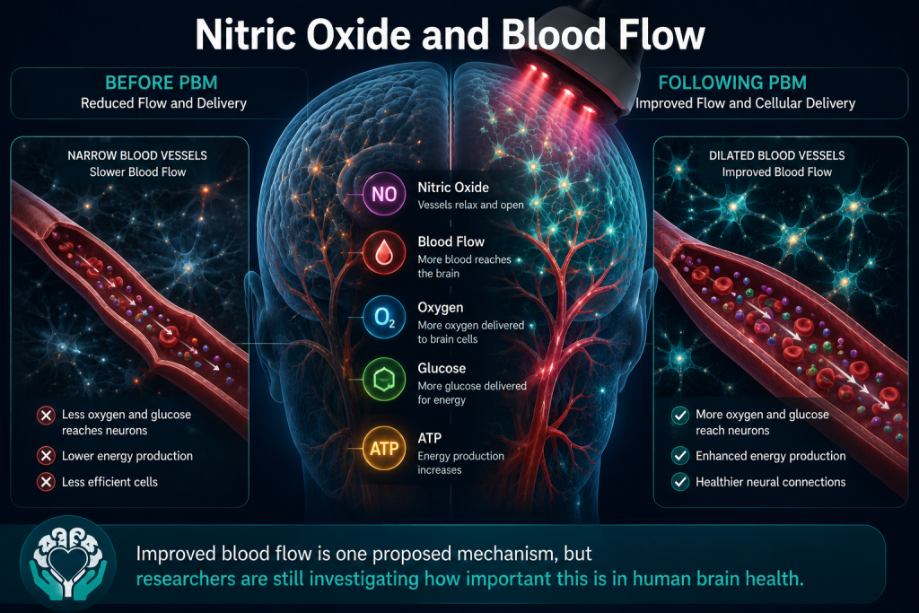
One particularly interesting molecule involved in this process is nitric oxide.
Despite its name, nitric oxide is not harmful.
In fact, it plays a vital role throughout the body.
Among its many jobs, nitric oxide helps blood vessels relax and widen.
Scientists believe that when red or near-infrared light interacts with mitochondria, nitric oxide may be released from cytochrome c oxidase.
This could have two important effects.
First, it may allow mitochondria to produce ATP more efficiently.
Second, it may help widen nearby blood vessels.
Imagine traffic building up on a busy motorway.
If extra lanes suddenly opened, traffic would begin flowing more freely.
Researchers think nitric oxide may act in a similar way by allowing more oxygen-rich blood to reach active tissues.
Several human studies have reported changes in cerebral blood flow and oxygenation following transcranial photobiomodulation, although the exact mechanisms responsible are still being investigated.
Step 4: Improving Blood Flow to the Brain
The brain depends on a continuous supply of oxygen and glucose.
Even small reductions in blood flow can affect how well neurons work.
Researchers have found evidence that photobiomodulation may temporarily improve blood flow to certain areas of the brain.
This doesn't mean new blood vessels suddenly appear.
Instead, existing blood vessels may become slightly more efficient at delivering oxygen and nutrients where they are needed.
You can think of it like improving traffic flow through an existing road network rather than building entirely new roads.
Better blood flow may help support normal brain function, particularly in areas under metabolic stress.
However, scientists are still investigating how long these effects last and whether they translate into meaningful improvements for people living with neurological conditions.
Step 5: Triggering Cellular Communication
Cells don't work in isolation.
They are constantly sending messages to one another.
Researchers believe photobiomodulation may influence this communication by activating cell signalling pathways.
Think of these pathways as the cell's internal email system.
When one part of the cell detects a change, messages are sent throughout the cell telling different proteins what to do next.
Some messages encourage repair.
Others help produce protective proteins.
Some switch inflammation on.
Others switch it down again.
Scientists have identified changes in the activity of numerous genes and signalling molecules following photobiomodulation in laboratory studies.
Importantly, the light itself does not change a person's DNA.
Instead, it may influence which existing genes become more or less active for a period of time, much like adjusting the volume on different instruments in an orchestra.
Step 6: Reducing Inflammation
Inflammation is one of the brain's natural defence mechanisms.
Immediately after an injury, it helps remove damaged cells and begin the repair process.
The problem comes when inflammation becomes excessive or continues for too long.
Chronic inflammation has been linked with traumatic brain injury, Alzheimer's disease, Parkinson's disease and many other neurological conditions.
Laboratory studies suggest photobiomodulation may help regulate inflammatory responses by influencing immune cells and chemical messengers involved in inflammation.
This does not mean inflammation is eliminated.
Nor does it mean PBM acts like an anti-inflammatory drug.
Instead, researchers believe it may help restore a healthier balance between helpful and harmful inflammatory responses.
Exactly how important this effect is in humans remains one of the major questions current clinical trials are trying to answer.
Step 7: Protecting Cells from Oxidative Stress
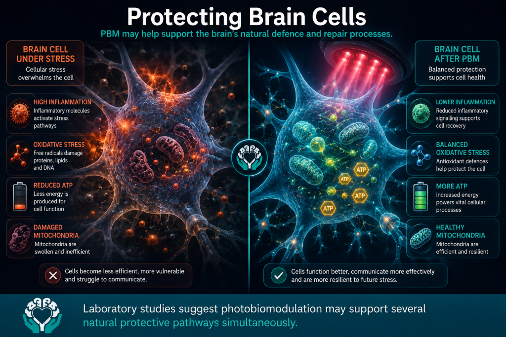
Every time our cells produce energy, they also generate tiny unstable molecules known as reactive oxygen species, or ROS.
In small amounts these molecules are completely normal.
In fact, they help cells communicate.
Problems arise when too many are produced.
This creates oxidative stress, which can damage proteins, fats and DNA if the body's natural antioxidant systems become overwhelmed.
Researchers have found that photobiomodulation may influence this balance.
Rather than eliminating ROS altogether—which would actually be harmful—it appears to encourage the small signalling amounts needed for healthy cell communication while also supporting the cell's own antioxidant defences.
This delicate balance may be one reason PBM has attracted interest across so many areas of neuroscience.
Step 8: Supporting Neuroplasticity
One of the brain's most remarkable abilities is neuroplasticity—its capacity to adapt, reorganise and form new connections.
Every time you learn something new...
Recover from an injury...
Or practise a new skill...
Your brain is physically changing.
Researchers have found evidence in laboratory studies that photobiomodulation may increase proteins involved in learning, memory and synaptic communication.
Some animal studies have also suggested it may encourage the growth of new neuronal connections and support the survival of existing brain cells after injury.
Human evidence, however, is much more limited.
Scientists do not yet know whether these laboratory findings translate into significant improvements in long-term brain repair in people living with traumatic brain injury or dementia.
Step 9: Neuroprotection – Helping Cells Cope with Stress
Perhaps the most exciting possibility is something researchers call neuroprotection.
Rather than repairing damage that has already occurred, neuroprotection means helping brain cells survive future stress.
Laboratory studies suggest photobiomodulation may strengthen several of the brain's natural protective systems at the same time.
These include:
- Supporting mitochondrial function.
- Improving blood flow.
- Regulating inflammation.
- Reducing oxidative stress.
- Encouraging cellular repair pathways.
Together, these effects may make neurons more resilient.
However, this remains one of the biggest unanswered questions in the field.
Scientists still need to determine whether these biological changes lead to meaningful clinical improvements for patients.
The Important Thing to Remember
One of the biggest misconceptions about photobiomodulation is that scientists know exactly how it works.
They don't.
Researchers agree that red and near-infrared light can influence cellular biology.
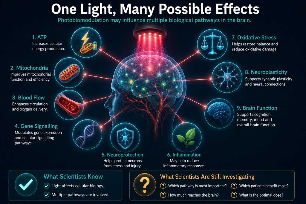
They have observed changes in:
- Energy production.
- Blood flow.
- Brain oxygenation.
- Inflammation.
- Cellular signalling.
- Brain imaging.
But the precise biological pathway—or pathways—responsible is still being investigated.
Cytochrome c oxidase remains the leading explanation, but it is unlikely to be the only one.
As our understanding of neuroscience grows, the story of photobiomodulation is likely to become even more complex—and even more interesting.
One of the biggest questions in photobiomodulation research—and one scientists are working hard to answer.
If someone told you that shining a light on your head could affect your brain, your first reaction would probably be:
"But surely my skull blocks the light?"
It's an excellent question.
In fact, it's one of the first questions scientists asked themselves when photobiomodulation research began.
The answer isn't simply yes or no.
Some light does reach the brain.
Most of it doesn't.
The real scientific question isn't whether light reaches the brain—it's whether the small amount that does is enough to produce meaningful biological effects.
This remains one of the most important areas of ongoing photobiomodulation research.
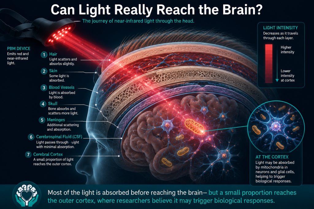
The Brain Is Protected by Several Layers
Our brains are designed to be well protected.
Before light can reach brain tissue, it must first travel through several different layers.
These include:
- Hair (if present)
- The skin of the scalp
- Blood vessels beneath the skin
- Connective tissue
- The skull
- The protective membranes surrounding the brain
- Cerebrospinal fluid (CSF)
- Finally, the outer layer of the brain itself, known as the cerebral cortex
Each of these layers absorbs, reflects or scatters some of the incoming light.
Imagine trying to shine a torch through several thick curtains.
Each curtain blocks a little more of the light.
By the time the light reaches the other side, it is much weaker than when it first started.
The same thing happens inside the head.
Every layer reduces the amount of light that continues towards the brain.
Not all colours of light behave in the same way.
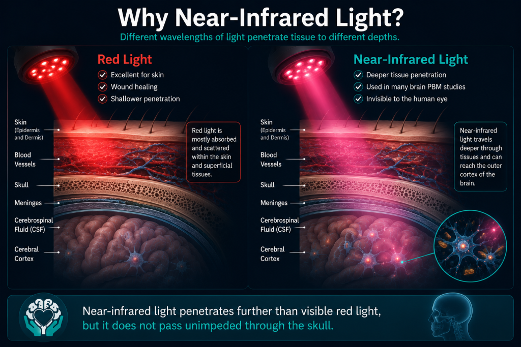
Visible red light is excellent for treating tissues close to the surface, such as the skin.
Near-infrared light, however, has an important advantage.
Although we cannot see it, these longer wavelengths can travel further through biological tissues before being absorbed.
Think of it like fog lights on a car.
In thick fog, some colours of light scatter quickly, making it difficult to see ahead.
Other wavelengths penetrate much further, allowing you to see more clearly.
Near-infrared light behaves in a similar way inside the body.
It doesn't pass straight through the skull, but it generally penetrates deeper than visible red light, making it more suitable for researchers investigating the brain.
This is why many transcranial photobiomodulation devices use wavelengths around 810 nanometres, although researchers continue to investigate other wavelengths and combinations.
How Much Light Actually Reaches the Brain?
This is where things become particularly interesting.
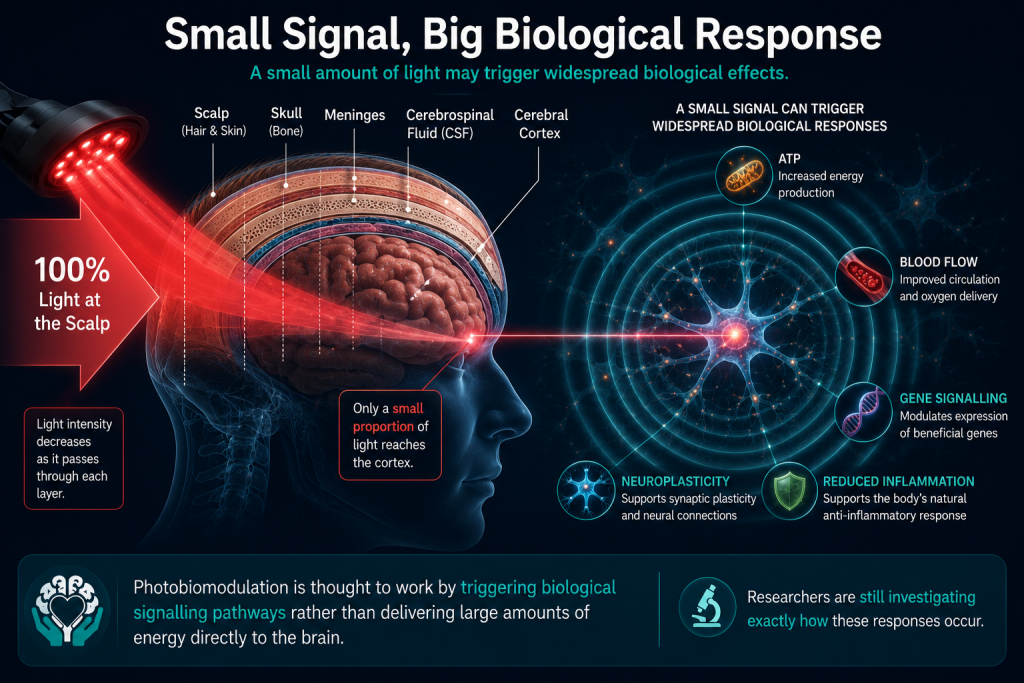
Scientists know that only a small percentage of the light applied to the scalp reaches the surface of the brain.
In one frequently cited study related to acute traumatic brain injury, researchers estimated that approximately 3% of the 810-nanometre light delivered at the scalp may have reached the outer surface of the cerebral cortex under the specific experimental conditions used.
At first glance, that might not sound very impressive.
After all...
97% didn't reach the brain.
Doesn't that mean the treatment can't work?
Not necessarily.
That 3% estimate came from one particular wavelength, one specific device and one experimental model. Other laboratory studies have reported different—and sometimes lower—levels of light transmission depending on factors such as skull thickness, wavelength, anatomy and the equipment being used. There is no single percentage that applies to every device or every person.
One of the biggest misconceptions about photobiomodulation is the idea that large amounts of light must reach the brain for it to have any effect.
Biology doesn't always work that way.
Consider a thermostat in your home.
You don't need enormous force to change the temperature.
A tiny movement of the dial can activate an entire heating system.
Or think about a light switch.
Pressing it requires almost no effort, yet it controls all the lights in a room.
Cells behave in a similar way.
Researchers believe photobiomodulation acts more like a biological signal than an energy source.
The light isn't trying to "power" the brain directly.
Instead, it may trigger natural cellular processes that amplify the original signal, allowing relatively small amounts of light to produce measurable biological responses.
This is one reason scientists remain interested in photobiomodulation despite the relatively small amount of light that reaches brain tissue.
Can Light Reach the Whole Brain?
This is where caution is needed.
Many advertisements suggest that red light "bathes the entire brain" or reaches deep structures such as the hippocampus with ease.
The scientific evidence is much more cautious.
Researchers generally agree that the greatest amount of light reaches the outer layers of the brain, particularly the cerebral cortex directly beneath the light source.
Much less light is expected to reach deeper brain structures because every additional millimetre of tissue absorbs and scatters more photons.
That doesn't necessarily mean deeper regions are completely unaffected.
Scientists have proposed several possible explanations, including:
- Networks of connected neurons transmitting biological changes from one brain region to another.
- Changes in blood flow affecting wider areas of the brain.
- Chemical signalling between cells.
- Systemic biological responses beyond the immediate area exposed to light.
At present, however, researchers are still investigating which of these mechanisms are most important.
What About Intranasal Light Therapy?
Some photobiomodulation systems also include a small light applicator that fits inside one nostril.
The idea is that the nasal cavity contains thinner layers of bone and tissue than the skull, potentially allowing light to reach nearby structures more easily.
Some researchers have suggested this may provide an additional pathway for delivering light.
However, the scientific evidence is still evolving.
Most studies using intranasal photobiomodulation combine it with light applied to the scalp, making it difficult to determine exactly which component is responsible for any observed effects.
Further research is needed before firm conclusions can be drawn about the contribution of intranasal therapy alone.
Why Device Design Matters
This is also why not all photobiomodulation devices are the same.
Two devices may both advertise "810 nanometre light," yet still deliver very different amounts of light to the brain.
Factors that influence how much light reaches brain tissue include:
- Wavelength.
- Light intensity (irradiance).
- Total energy delivered.
- Continuous or pulsed light.
- Size of the light source.
- How closely the device contacts the scalp.
- Hair thickness.
- Skin colour.
- Skull thickness.
- Individual anatomy.
This is one reason researchers cannot assume that results seen with one scientifically tested device automatically apply to every commercially available red-light helmet, panel or handheld device.
The entire treatment protocol—not just the colour of the light—matters.
The Bottom Line
Scientists no longer question whether some light can reach the brain.
Laboratory studies and sophisticated imaging techniques have shown that it can.
The much bigger question is:
Is the amount that reaches the brain enough to produce meaningful improvements for people living with neurological conditions?
Early research suggests it may be.
Studies have reported changes in brain metabolism, blood flow, oxygenation and MRI measurements following transcranial photobiomodulation.
However, researchers are still determining how these biological changes relate to improvements in symptoms, thinking, memory and long-term recovery.
That's why larger clinical trials remain so important.
The History of Photobiomodulation
From an accidental discovery to one of the most exciting areas of modern neuroscience.
The idea that light might help heal the body isn't new.
In fact, doctors have been investigating the medical use of light for well over a century.
What has changed is our understanding of how light interacts with living cells.
Today, photobiomodulation is being studied in some of the world's leading neuroscience laboratories as a potential treatment for traumatic brain injury, Alzheimer's disease, Parkinson's disease and other neurological conditions.
But the journey to get here has been long—and it all began with an unexpected observation.
The First Medical Use of Light
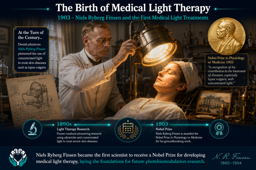
Long before lasers or LED devices existed, doctors had already noticed that certain types of light appeared to influence health.
In the late 1800s, Danish physician Dr Niels Ryberg Finsen became fascinated by the effects of light on disease.
At the time, tuberculosis was one of the world's deadliest illnesses. One form of the disease, known as lupus vulgaris, caused painful tuberculosis infections of the skin.
Finsen developed special lamps that concentrated particular wavelengths of light onto these skin lesions.
Remarkably, many patients improved.
His work demonstrated something revolutionary for the time:
Light could produce real biological effects inside living tissue.
For this pioneering research, Finsen was awarded the 1903 Nobel Prize in Physiology or Medicine, becoming one of the first scientists recognised for developing light-based medical treatments.
Although his work had nothing to do with the brain, it laid the foundations for everything that would follow.
For the first time, medicine had proof that carefully controlled light could influence biology without surgery or drugs.
Fast forward more than sixty years.
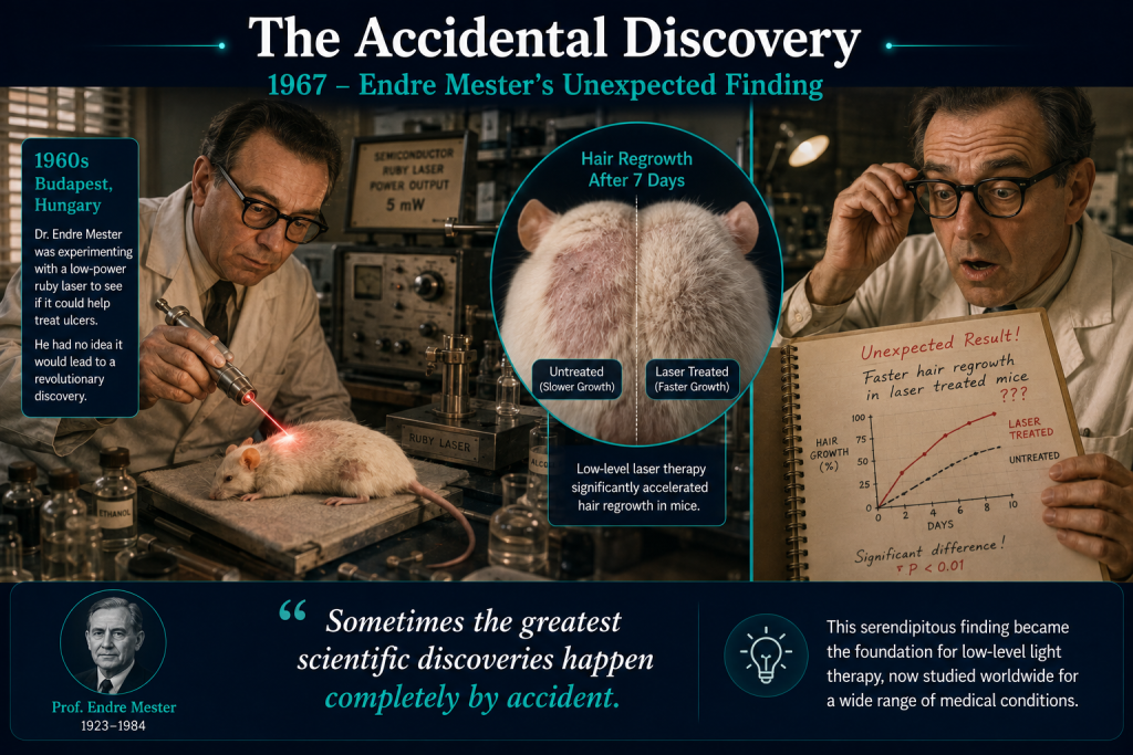
In the 1960s, lasers had recently been invented, and scientists were excited about their potential medical uses.
One of those researchers was Hungarian physician Professor Endre Mester.
His team wanted to investigate whether lasers could be used to treat cancer.
To test the idea, they shaved patches of fur from laboratory mice and exposed them to a low-powered ruby laser.
The researchers expected to see damage.
Instead...
Something completely unexpected happened.
The mice didn't develop cancer.
Their skin wasn't burned.
In fact, the hair on the treated areas appeared to grow back faster than the untreated areas.
Rather than damaging tissue, the low-powered laser seemed to be encouraging it to heal.
It was an accidental discovery—but one that would change medical research forever.
Professor Mester went on to investigate whether low-powered light could improve wound healing and tissue repair in both animals and people.
His studies suggested that light could stimulate normal biological processes without generating enough heat to burn or destroy tissue.
Today, many scientists consider these experiments to mark the birth of modern photobiomodulation research.
From "Cold Lasers" to Photobiomodulation
As research expanded during the 1970s, 1980s and 1990s, scientists used many different names for this emerging therapy.
You may still come across terms such as:
- Cold Laser Therapy
- Low-Level Laser Therapy (LLLT)
- Low-Power Laser Therapy
- Laser Biostimulation
- Red-Light Therapy
While these names describe similar ideas, they can also be confusing.
Not every treatment uses a laser.
Some use LEDs.
Some use red light.
Others use near-infrared light.
To bring consistency to the field, international experts eventually agreed on a new name:
Photobiomodulation Therapy, or simply PBM.
The word may sound complicated, but it actually describes the process quite well:
- Photo = light
- Bio = living cells
- Modulation = influencing or adjusting biological activity
In other words:
Using light to influence how living cells function.
Unlike laser surgery, PBM does not aim to destroy tissue.
Instead, it aims to encourage the body's own natural cellular processes.
The Rise of LED Technology
One of the biggest changes in photobiomodulation over the past two decades has been the move away from lasers alone.
Early research relied mainly on medical lasers because they were the only technology capable of delivering precise wavelengths of light.
However, engineers soon realised that light-emitting diodes (LEDs) could also produce the same biologically active wavelengths.
LEDs offered several advantages.
They:
- Cover larger treatment areas.
- Produce much less heat.
- Are generally less expensive.
- Can be built into wearable devices.
- Are easier to use outside hospital settings.
Today, many of the most widely studied transcranial photobiomodulation systems use arrays of carefully positioned LEDs rather than a single laser beam.
This has made it possible to develop lightweight headsets and helmets designed specifically for research into brain disorders.
Bringing Light to the Brain
For many years, photobiomodulation research focused mainly on wound healing, sports injuries, muscle recovery and pain management.
As scientists learned more about mitochondria and cellular energy production, an intriguing question emerged.
Could the same biological effects help brain cells?
Laboratory studies began suggesting that red and near-infrared light might influence:
- ATP production.
- Blood flow.
- Inflammation.
- Oxidative stress.
- Neuronal survival.
These discoveries sparked a completely new field of research:
Transcranial Photobiomodulation (tPBM)—the application of light to the head with the aim of influencing brain function.
Today, researchers around the world are investigating tPBM for conditions including:
- Traumatic brain injury (TBI).
- Persistent post-concussion symptoms.
- Alzheimer's disease.
- Mild cognitive impairment.
- Parkinson's disease.
- Stroke.
- Depression.
Although many studies remain in the early stages, the number of clinical trials has increased dramatically over the last decade.
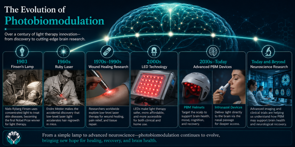
Modern Helmets and Intranasal Devices
Modern photobiomodulation systems look very different from the laboratory lasers used in the 1960s.
Many research devices now resemble lightweight helmets or headbands lined with dozens of LEDs.
These are designed to deliver carefully controlled doses of red or near-infrared light to different regions of the scalp.
Some systems also include intranasal light applicators.
These small devices fit inside one nostril and deliver near-infrared light into the nasal cavity.
Researchers are investigating whether this route may provide an additional way of influencing nearby tissues or biological signalling pathways.
However, scientists are still studying exactly how much light reaches the brain through this route and what contribution, if any, intranasal therapy makes to overall treatment effects.
Most clinical studies combine scalp and intranasal treatment, making it difficult to separate the effects of each individually.
The Rise of Consumer Devices
As public interest in photobiomodulation has grown, so too has the number of products available online.
Today it's possible to buy:
- Red-light panels.
- Handheld light devices.
- Wearable helmets.
- Caps.
- Face masks.
- Intranasal devices.
Some are based on technologies similar to those being studied in clinical research.
Others have little published evidence behind them.
This is an important distinction.
A device advertised as "red light therapy" is not automatically supported by scientific evidence simply because it uses a similar colour of light.
The effectiveness of photobiomodulation depends on many factors, including:
- The wavelength used.
- The amount of energy delivered.
- The treatment schedule.
- Whether the light reaches the intended tissue.
- The condition being treated.
Evidence gathered using one carefully designed research device cannot automatically be applied to every commercial product on the market.
A Field That Is Still Evolving
Photobiomodulation has travelled an extraordinary journey.
What began as an unexpected observation in laboratory mice has developed into an international field of research involving neuroscientists, physicists, engineers and clinicians.
Today, hundreds of scientific papers have explored how red and near-infrared light may influence cells.
Yet despite decades of research, many important questions remain unanswered.
Scientists are still working to determine:
- Which wavelengths work best.
- The ideal treatment dose.
- How often treatment should be given.
- Which patients are most likely to benefit.
- Whether changes seen in laboratory studies translate into meaningful improvements in people's lives.
As with many promising medical technologies, the science continues to evolve.
The history of photobiomodulation reminds us that some of medicine's greatest discoveries begin not with certainty—but with curiosity.
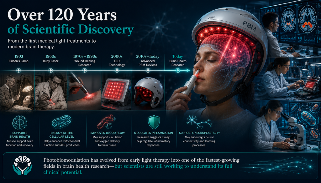
Why Are Scientists Interested in Brain Injury?
Understanding why photobiomodulation has become one of the most promising areas of traumatic brain injury research.
When someone experiences a concussion or traumatic brain injury (TBI), the damage doesn't stop at the moment of impact.
For many years, scientists believed that the injury itself was the main problem.
Today, we know that's only part of the story.
The initial blow to the head is often just the first chapter.
Over the hours, days and sometimes weeks that follow, the brain undergoes a series of complex biological changes that can cause additional damage. Researchers call this secondary injury.
Understanding this process has completely changed how scientists think about treating brain injuries.
Rather than focusing only on the impact itself, researchers are now asking an important question:
Can we help the brain protect and repair itself during the critical hours and days after injury?
This is one reason photobiomodulation has attracted so much scientific interest.
Unlike treatments that target a single symptom, researchers believe PBM may influence several of the biological processes involved in secondary injury—including energy production, inflammation and cellular repair. While this remains an area of active research, it offers a very different approach to supporting recovery.
The First Injury Is Only the Beginning
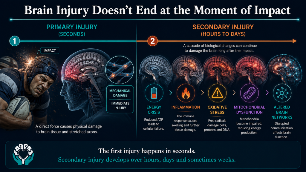
Imagine dropping your mobile phone onto a hard floor.
The crack in the screen happens instantly.
That's the primary injury.
Now imagine that after the fall, the battery begins overheating, the software starts crashing and internal components slowly stop working over the next few hours.
Those later problems weren't caused by a second drop.
They happened because the original damage triggered a chain reaction.
Something very similar happens inside the brain.
The primary injury occurs at the moment of impact.
This may involve stretching of nerve fibres (axons), bruising, bleeding or direct damage to brain tissue.
But after that initial trauma, a second wave of biological changes begins.
These changes are known collectively as secondary injury, and in many cases they may contribute more to long-term symptoms than the original impact itself.
This concept is central to modern brain injury research and was discussed in our previous article, What Actually Happens Inside the Brain During a Concussion? There, we explored how rotational forces stretch delicate nerve fibres and trigger a cascade of biochemical events that can continue long after the initial blow.
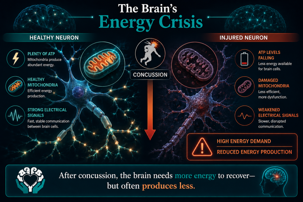
One of the earliest events after a concussion is something scientists call the neurometabolic or energy crisis.
Immediately after the injury, millions of brain cells lose their normal electrical balance.
To restore this balance, they suddenly need enormous amounts of energy.
It's rather like a city after a major power cut.
Every engineer is working overtime to restore electricity, repair damaged infrastructure and get essential services running again.
The demand for power skyrockets.
The problem is that the brain's power stations—its mitochondria—are often working less efficiently at exactly the same time.
This creates a dangerous mismatch.
The brain needs more energy than ever before, but its ability to produce that energy has been reduced.
Researchers believe this energy crisis may contribute to many of the symptoms experienced after concussion, including:
- Brain fog.
- Mental fatigue.
- Slower thinking.
- Poor concentration.
- Memory problems.
- Difficulty processing information.
For most people, the brain gradually restores its normal energy balance over days or weeks.
For others, particularly after repeated concussions or more severe traumatic brain injuries, recovery may take much longer.
Mitochondria Under Pressure
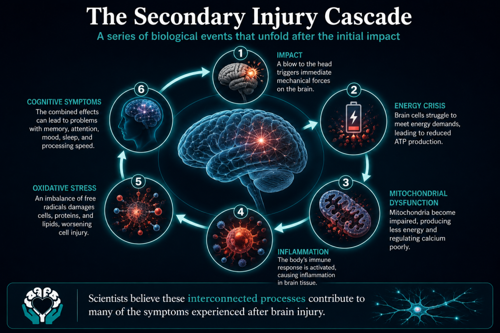
As we explored earlier, mitochondria are the tiny structures responsible for producing ATP—the energy that powers every brain cell.
Following a traumatic brain injury, these microscopic power stations can become disrupted.
Scientists have observed changes in:
- Mitochondrial respiration (how efficiently mitochondria produce energy).
- Calcium regulation.
- Oxidative stress.
- The balance between energy production and energy demand.
Rather than producing ATP efficiently, injured mitochondria may begin generating more harmful reactive oxygen species while producing less usable energy.
Imagine trying to run an entire hospital using emergency generators.
The lights stay on—but not everything works as efficiently as it should.
Researchers believe something similar may happen inside injured neurons.
This mitochondrial dysfunction has become one of the major reasons scientists are investigating therapies such as photobiomodulation that aim to support cellular energy production rather than simply treating symptoms.
When Inflammation Doesn't Switch Off
Inflammation often gets a bad reputation.
In reality, it's one of the body's most important defence mechanisms.
Immediately after an injury, specialised immune cells move into the damaged area.
Their job is to:
- Remove damaged tissue.
- Clear away debris.
- Fight infection.
- Begin the healing process.
Without inflammation, recovery would be impossible.
The problem comes when this response becomes excessive or continues for too long.
Following traumatic brain injury, some inflammatory processes can remain active long after the initial injury has healed.
Researchers believe prolonged inflammation may contribute to ongoing symptoms and may also influence the long-term health of neurons.
Laboratory studies suggest photobiomodulation may help regulate inflammatory signalling rather than simply suppressing it.
This distinction is important.
Scientists are not trying to eliminate inflammation completely.
Instead, they hope to encourage a more balanced response that supports healing while reducing unnecessary damage.
Whether this translates into meaningful clinical improvements for patients is still being investigated.
Oxidative Stress – When Cells Become Overloaded
As injured brain cells struggle to restore normal function, they often produce increased amounts of unstable molecules called reactive oxygen species (ROS).
In small amounts, ROS are completely normal.
They even help cells communicate with one another.
However, when too many are produced, they can overwhelm the brain's natural antioxidant defences.
This leads to oxidative stress.
You can think of oxidative stress a little like rust developing on metal.
A small amount of wear is part of everyday life.
But if damage accumulates faster than repairs can be made, the structure gradually begins to weaken.
Researchers believe oxidative stress may contribute to ongoing cellular damage following concussion and traumatic brain injury.
Animal and laboratory studies suggest photobiomodulation may help support the cell's natural antioxidant systems, although scientists are still determining how important this effect is in human patients.
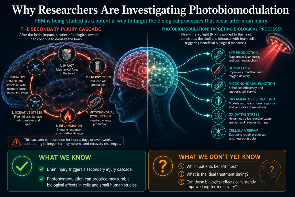
One of the biggest challenges in treating traumatic brain injury is that there isn't just one problem to fix.
The injured brain is dealing with several biological challenges at the same time.
These include:
- Reduced ATP production.
- Mitochondrial dysfunction.
- Changes in blood flow.
- Inflammation.
- Oxidative stress.
- Disrupted communication between neurons.
Most medicines are designed to target a single pathway.
Photobiomodulation is different.
Researchers believe it may influence multiple interconnected biological systems simultaneously, potentially helping the brain's own repair processes during this vulnerable period.
That's an exciting idea.
But it's important to keep it in perspective.
While laboratory research and early clinical trials have reported encouraging biological changes, scientists are still working to determine whether these changes consistently lead to better long-term recovery, improved cognition and enhanced quality of life for people living with brain injuries.
That is exactly what ongoing clinical trials aim to answer.
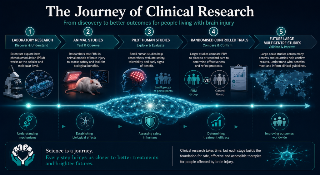
What Does the Research Show After Brain Injury?
Promising early results—but are we ready to call photobiomodulation an effective treatment for traumatic brain injury?
Photobiomodulation has generated enormous interest as a potential therapy for traumatic brain injury (TBI).
Laboratory studies have shown that red and near-infrared light can influence many of the biological processes involved in brain injury, including mitochondrial function, inflammation, blood flow and cellular signalling.
But an important question remains.
Do these biological changes actually help people recover?
That's where human clinical trials become essential.
While the science behind photobiomodulation is compelling, researchers need to demonstrate that improvements seen in laboratory experiments translate into meaningful benefits for patients—such as better memory, clearer thinking, reduced symptoms and improved quality of life.
So far, the evidence is encouraging, but it is also limited.
Let's look at what the research tells us.
Acute Traumatic Brain Injury
Can Photobiomodulation Help in the First Few Days After Injury?
The hours immediately after a traumatic brain injury are incredibly important.
This is when the brain is experiencing the secondary injury cascade we explored earlier—an energy crisis, inflammation, oxidative stress and disrupted cellular function.
Researchers believe this may be the ideal time to intervene.
If photobiomodulation can support brain cells while these biological processes are unfolding, it might help reduce some of the damage caused by secondary injury.
This theory led to one of the most important human studies published so far.
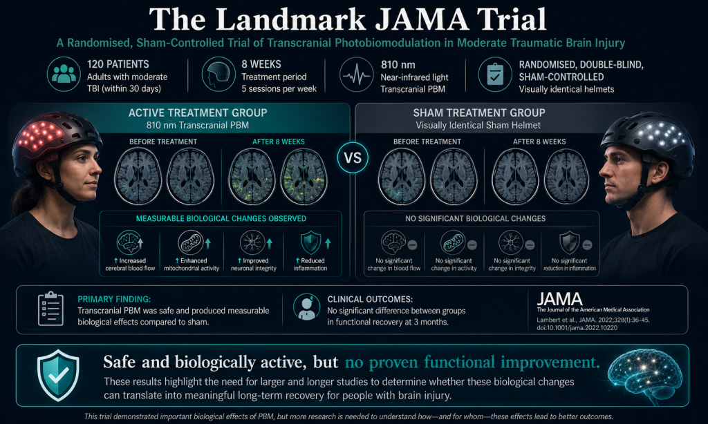
In 2020, researchers published the first randomised, sham-controlled clinical trial investigating transcranial photobiomodulation in people with moderate traumatic brain injury.
Published in JAMA Network Open, the study included 68 patients who received either active near-infrared light therapy or a sham (placebo) treatment within 72 hours of injury.
Participants received three 20-minute treatment sessions using 810-nanometre light delivered through the scalp.
The primary aim of the study was not to prove that patients recovered faster.
Instead, researchers wanted to answer two important questions:
- Was the treatment safe?
- Could it produce measurable biological effects inside the injured brain?
The answer to both questions was encouraging.
Researchers found no treatment-related serious adverse events, suggesting that short-term treatment appeared to be well tolerated.
More importantly, advanced MRI scans detected measurable differences between the brains of participants who received active treatment and those who received sham therapy.
Changes Seen on Brain MRI
Rather than relying only on symptom questionnaires, researchers used sophisticated diffusion MRI techniques to examine the brain's white matter.
White matter acts like the brain's communication network, allowing different regions to exchange information.
Following traumatic brain injury, these connections can become disrupted.
The MRI scans suggested that participants receiving photobiomodulation showed changes in white matter that may indicate a biological response to treatment.
This was an exciting finding because it suggested the light was doing something measurable inside the injured brain.
However, it's important to understand what this doesn't mean.
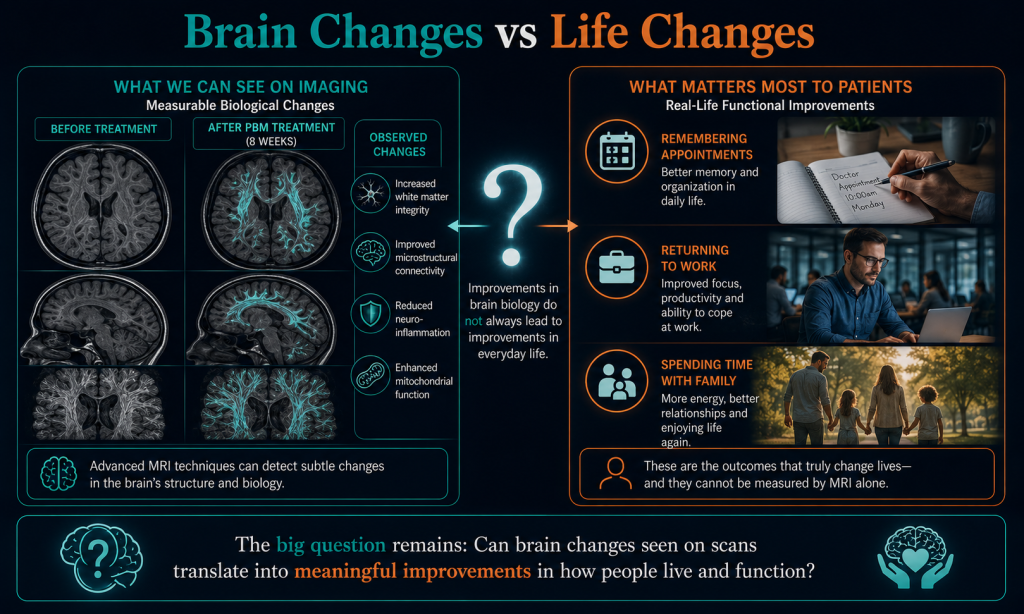
An MRI change is not the same as proving someone thinks more clearly, remembers more information or returns to work sooner.
Brain imaging helps researchers understand biology.
Patients care about outcomes that affect everyday life.
Connecting these two remains one of the biggest challenges in brain injury research.
Did Patients Actually Recover Better?
Interestingly, people who received photobiomodulation reported slightly lower symptom scores three months later than those receiving sham treatment.
However, these improvements were not statistically strong enough to prove that the treatment itself caused the difference.
The study simply wasn't large enough to answer that question.
Instead, the researchers concluded that the trial demonstrated:
- The treatment appeared safe.
- The brain showed measurable biological responses.
- Larger clinical trials were now justified.
In science, this was an important step forward—but it was not proof that photobiomodulation improves recovery after traumatic brain injury.
Persistent Post-Concussion Symptoms
What About People Who Continue to Have Symptoms?
Most people recover from concussion within days or weeks.
Unfortunately, some continue experiencing symptoms for months or even years.
These may include:
- Brain fog.
- Fatigue.
- Headaches.
- Poor concentration.
- Memory problems.
- Sleep disturbance.
- Sensitivity to light and noise.
Researchers have become increasingly interested in whether photobiomodulation might help this group.
Several small pilot studies have now investigated this possibility.
Early Pilot Studies
One pilot study published in 2024 followed people living with persistent cognitive symptoms after mild traumatic brain injury.
Some participants reported improvements in:
- Cognitive symptoms.
- Sleep.
- Overall wellbeing.
However, objective neuropsychological testing showed improvement in only a small number of participants.
The researchers themselves highlighted several important limitations.
Participants were taking different medications.
Some were receiving other therapies.
The study involved relatively few people.
Without a larger placebo-controlled trial, it was impossible to know how much of the improvement resulted from photobiomodulation itself.
The 2025 Randomised Crossover Study
A more recent study published in 2025 attempted to answer this question more rigorously.
Seventeen participants with mild traumatic brain injury received both active photobiomodulation and sham treatment at different times.
Researchers reported improvements following active treatment in several areas, including:
- Visual working memory.
- Verbal learning.
- Sleep.
- Pain.
- Post-concussion symptoms.
These findings are encouraging.
However, once again the study had important limitations.
The number of participants was very small.
Several comparisons between active and placebo treatment did not reach clear statistical significance.
Repeated cognitive testing may also have introduced learning effects.
The authors concluded that their findings support the need for larger studies rather than proving effectiveness.
Chronic Traumatic Brain Injury
Can Photobiomodulation Help Years After Injury?
Some of the earliest photobiomodulation studies focused on people living with longstanding brain injuries.
Many of these reports describe individuals who experienced improvements in attention, memory, executive function, sleep or quality of life after treatment.
At first glance, these results appear extremely encouraging.
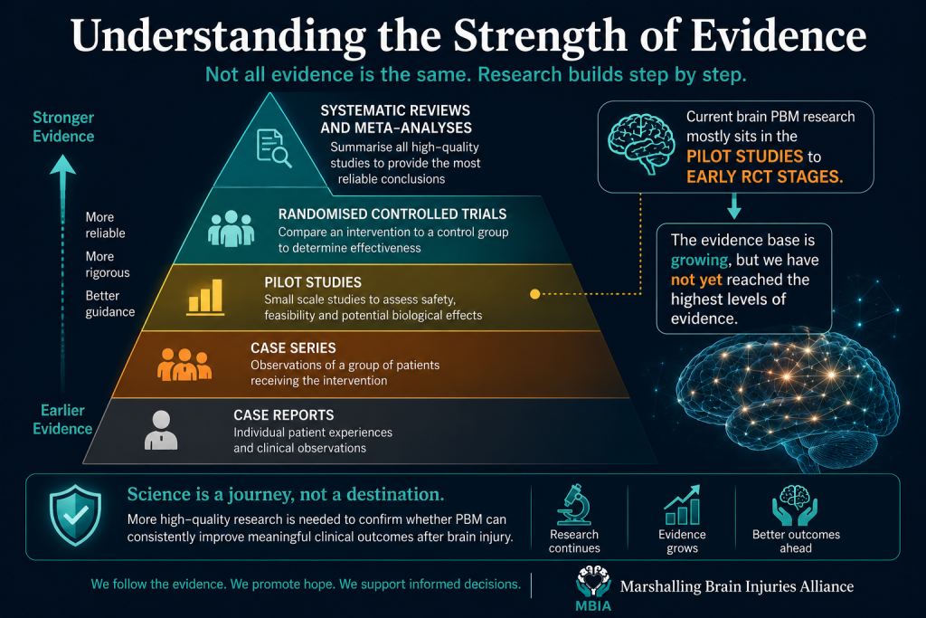
However, most of these publications were case reports or small case series.
What Is a Case Report?
A case report simply describes what happened to one individual or a small group of patients.
For example:
A clinician treats a patient with photobiomodulation.
The patient reports feeling better.
The doctor publishes the experience.
Case reports are valuable because they often generate new research ideas.
Many important medical discoveries began with individual patients.
However, they cannot prove that a treatment works.
Why?
Because there are many other possible explanations.
The patient might have improved naturally.
They may have been receiving rehabilitation at the same time.
They may have expected the treatment to help, producing a placebo effect.
Or the improvement may simply reflect normal fluctuations in symptoms.
Without a comparison group receiving sham treatment, it's impossible to know for certain.
Why Higher-Quality Studies Matter
This is why scientists place greater weight on randomised, sham-controlled clinical trials.
These studies are specifically designed to answer one question:
Would patients have improved even if they hadn't received the active treatment?
By comparing active photobiomodulation with a carefully designed placebo treatment, researchers can separate genuine treatment effects from expectation, natural recovery and chance.
At present, there are relatively few high-quality human studies of photobiomodulation after traumatic brain injury.
Most involve small numbers of participants using different devices, wavelengths, treatment schedules and outcome measures.
This makes it difficult to compare results directly or determine which approach works best.
What Does the Overall Evidence Tell Us?
Taken together, the current research paints an encouraging—but cautious—picture.
Laboratory research provides a strong biological rationale for photobiomodulation.
Human studies suggest the treatment can produce measurable changes in brain biology.
Some patients have also reported improvements in symptoms and cognition.
However, the evidence remains too limited to conclude that photobiomodulation reliably improves recovery after traumatic brain injury.
Researchers now agree that the next step is larger, multicentre clinical trials involving hundreds of participants, longer follow-up periods and carefully standardised treatment protocols.
Only then will we know whether the biological effects seen in today's studies translate into meaningful improvements in people's lives.
What We Know
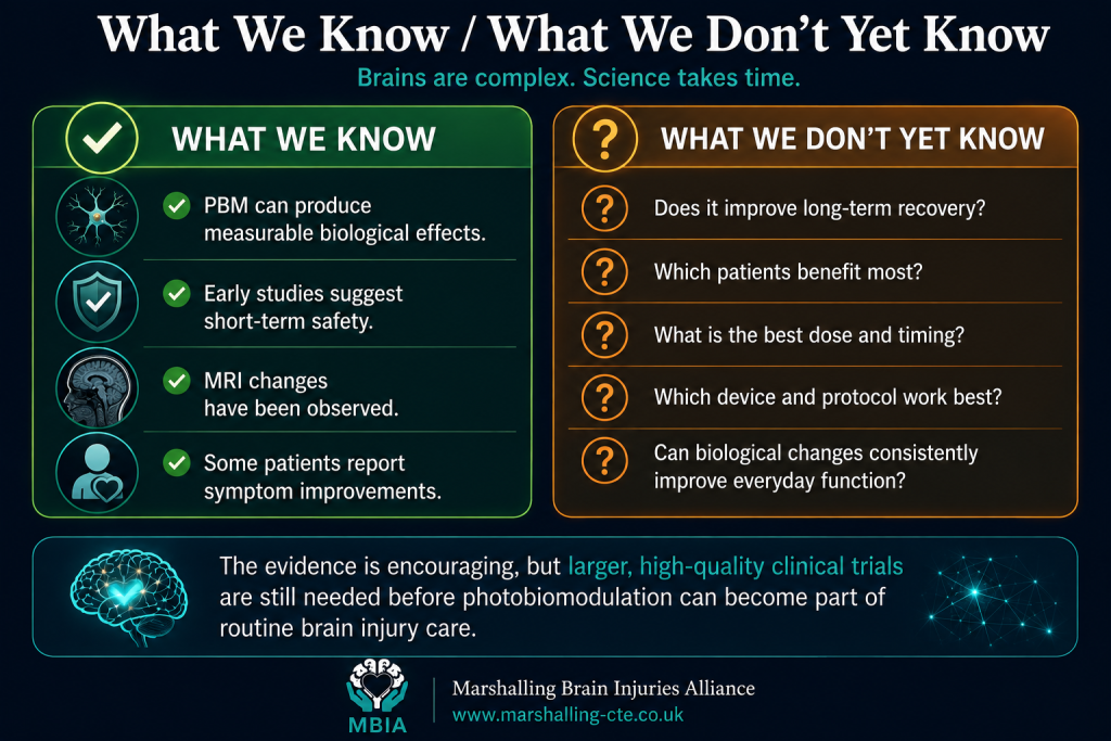
🟢 Researchers have demonstrated that transcranial photobiomodulation can produce measurable biological changes in the injured brain.
🟢 Small clinical studies suggest the treatment is generally well tolerated over the short term.
🟢 Advanced MRI studies have shown changes in white matter and brain connectivity following treatment.
🟢 Some patients report improvements in cognitive symptoms, sleep, fatigue and quality of life.
🟢 Laboratory evidence strongly supports effects on mitochondrial function, blood flow and cellular signalling.
What We Don't Yet Know
🟠 Does photobiomodulation consistently improve long-term recovery after traumatic brain injury?
🟠 Which patients benefit most—mild, moderate or severe injuries?
🟠 What is the ideal wavelength, dose and treatment schedule?
🟠 How soon after injury should treatment begin?
🟠 Do MRI changes translate into meaningful improvements in everyday function, independence and quality of life?
These are exactly the questions that ongoing clinical trials around the world are now trying to answer.
Why Aren’t Scientists Certain Yet?
The research is promising—but photobiomodulation studies are not yet consistent enough to give simple answers.
When people read about photobiomodulation online, the treatment can sometimes sound far more established than it really is.
One article may report improved memory.
Another may describe changes on a brain scan.
A company may then advertise a device using phrases such as:
- “Boosts brain energy.”
- “Repairs damaged neurons.”
- “Improves cognition.”
- “Supports recovery after concussion.”
The problem is that scientific evidence does not become reliable simply because several small studies report encouraging results.
Researchers must be able to repeat those findings in larger groups, using carefully controlled methods, and show that the effects are meaningful in everyday life.
At present, PBM research faces several important challenges.
These do not mean the treatment is ineffective.
They mean scientists are still working out whether it works reliably, for whom, at what dose and under which conditions.
Many Studies Include Very Few People
One of the biggest problems is study size.
Some PBM studies include fewer than 20 participants.
Others describe only a handful of individual cases.
Small studies are useful in the early stages of research because they can help answer questions such as:
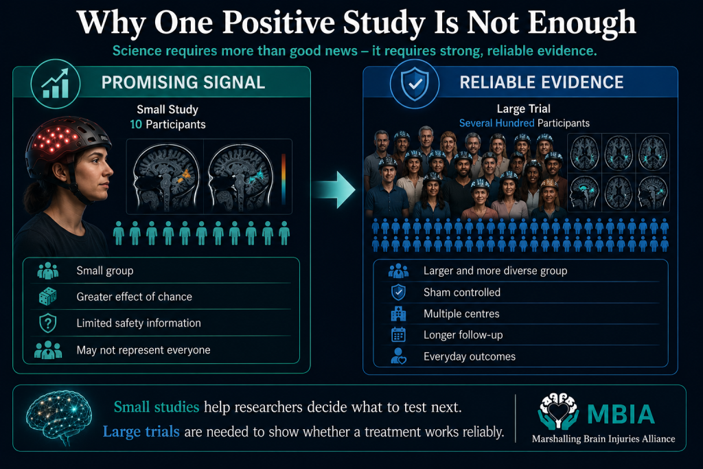
- Is the treatment practical?
- Does it appear safe?
- Can participants tolerate it?
- Are there early signs of biological activity?
- Which outcomes should future trials measure?
However, they are not very good at proving that a treatment works.
Imagine tossing a coin only four times.
You might get four heads in a row.
That result could look impressive, but it would not prove the coin is special. The result may simply have occurred by chance.
The same problem can happen in small clinical studies.
A few people may improve substantially, while others may not respond at all. With only a small number of participants, those individual responses can make the average result look much stronger than it would in a larger and more representative group.
Small studies are also less able to detect uncommon side effects or identify why some people respond differently from others.
This is why a positive pilot study should usually be treated as a reason to carry out a larger trial—not as proof that the treatment is effective.
Brain Injury and Dementia Are Not Single Conditions
Another challenge is that researchers are not always studying the same type of patient.
Traumatic brain injury can include:
- A recent concussion.
- Persistent post-concussion symptoms.
- Chronic mild TBI.
- Moderate or severe TBI.
- A single injury.
- Repeated head impacts.
- Injuries involving bleeding, swelling or diffuse axonal damage.
Alzheimer’s research may involve:
- Subjective cognitive concerns.
- Mild cognitive impairment.
- Early Alzheimer’s disease.
- Mild-to-moderate dementia.
- Mixed dementia.
- People with different genetic and vascular risk factors.
These groups are not interchangeable.
A treatment that helps someone during the first 72 hours after a moderate TBI may not have the same effect 10 years after repeated concussions.
Likewise, a therapy that improves a cognitive score in mild cognitive impairment may not help someone with advanced dementia.
When studies combine very different participants, it becomes difficult to know exactly who might benefit.
Different Devices May Deliver Very Different Treatments
The phrase photobiomodulation device covers a wide range of equipment.
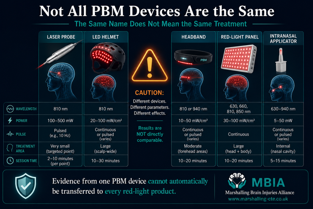
A study may use:
- A medical laser.
- An LED helmet.
- A flexible headset.
- A handheld probe.
- A large red-light panel.
- An intranasal applicator.
- A combination of scalp and intranasal light.
Two devices may look similar but deliver very different amounts of energy.
They may also differ in:
- The number of light sources.
- Their position on the head.
- How closely they contact the scalp.
- The treatment area.
- Whether the light is continuous or pulsed.
- The amount of heat produced.
- The accuracy of the stated output.
This means results from one device cannot automatically be applied to another.
It is similar to medicines.
Two tablets may both contain an active ingredient, but the dose, formulation and way the drug is absorbed can change the result.
With PBM, the complete device and treatment protocol matter—not simply whether the product produces red or near-infrared light.
Researchers Use Different Wavelengths
Wavelength describes the specific part of the light spectrum being used.
Red-light PBM often uses wavelengths in the visible red range, while brain studies commonly use near-infrared wavelengths because they tend to penetrate biological tissue more deeply.
However, studies do not all use the same wavelength.
Researchers have tested wavelengths including approximately:
- 630 nanometres.
- 660 nanometres.
- 808 nanometres.
- 810 nanometres.
- 850 nanometres.
- 1064 nanometres.
Some studies use one wavelength.
Others combine several.
This matters because different wavelengths may be absorbed by different cellular molecules and may travel through tissue differently.
A result using 810-nanometre light does not prove that 660-nanometre light—or every product advertised as red-light therapy—will have the same effect.
Scientists still need to determine whether there is one ideal wavelength or whether different conditions require different approaches.
The Dose Is Not Standardised
Even when two studies use the same wavelength, they may deliver very different doses.
Researchers must consider:
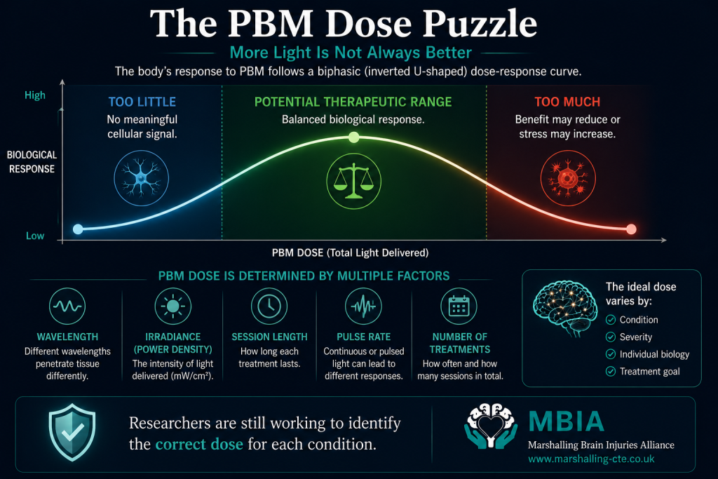
- Irradiance: how much power reaches a given area.
- Fluence: the total energy delivered to that area.
- Session length: how long the light is applied.
- Pulse pattern: whether the light is continuous or switches on and off.
- Duty cycle: how much of each pulse period the light remains active.
- Treatment area: how much of the scalp is exposed.
- Number of sessions: whether treatment is given once, several times or for months.
This creates one of the field’s biggest problems.
There is no universally accepted brain-PBM dose.
A treatment may be too weak to trigger a useful response.
Another may be within a helpful range.
A higher dose may be less effective.
This is known as a biphasic dose response.
An easy analogy is exercise.
Too little may not produce much benefit.
The right amount can improve health.
Too much may cause exhaustion or injury.
PBM may behave in a similar way.
More powerful light, longer sessions or more frequent treatment are not automatically better.
In some research, lower doses have produced more favourable results than higher doses on certain measures. This is why treatment parameters must be tested carefully rather than selected through guesswork.
Treatment Schedules Vary Widely
Studies also use very different treatment schedules.
A participant might receive:
- One session.
- Three sessions over several days.
- Treatment three times per week.
- Daily sessions.
- Six sessions per week.
- Treatment for four weeks.
- Treatment for eight or 12 weeks.
- Long-term home use.
The timing after brain injury may also be crucial.
Treatment started within hours of injury may aim to reduce the secondary injury cascade.
Treatment delivered years later may instead aim to influence chronic symptoms, plasticity or brain-network function.
Those are biologically different goals.
Until researchers compare schedules directly, they cannot confidently say:
- How quickly treatment should begin.
- How often it should be used.
- How long each session should last.
- How many weeks or months are needed.
- Whether benefits continue after treatment stops.
- Whether maintenance sessions are required.
Without those answers, it is difficult to build a reliable clinical protocol.
Hair, Skin and Skull Differences Affect the Dose
Even if every participant uses the same device, they may not receive the same amount of light at the brain.
Light can be influenced by:
- Hair thickness.
- Hair colour.
- Whether the device touches the scalp.
- Skin pigmentation.
- Skull thickness.
- Skull density.
- Head shape.
- The position of the light source.
A person with thick hair may absorb or scatter more light before it reaches the scalp.
A thicker skull may reduce transmission further.
This means the dose emitted by the device is not necessarily the dose reaching the cortex.
Scientists call this problem dosimetry—working out how much energy reaches the intended tissue.
Until studies measure and report this more consistently, comparing devices and trials will remain difficult.
Creating a Convincing Placebo Is Difficult
High-quality clinical trials usually compare the real treatment with a placebo, or sham treatment.
For PBM, this is harder than it sounds.
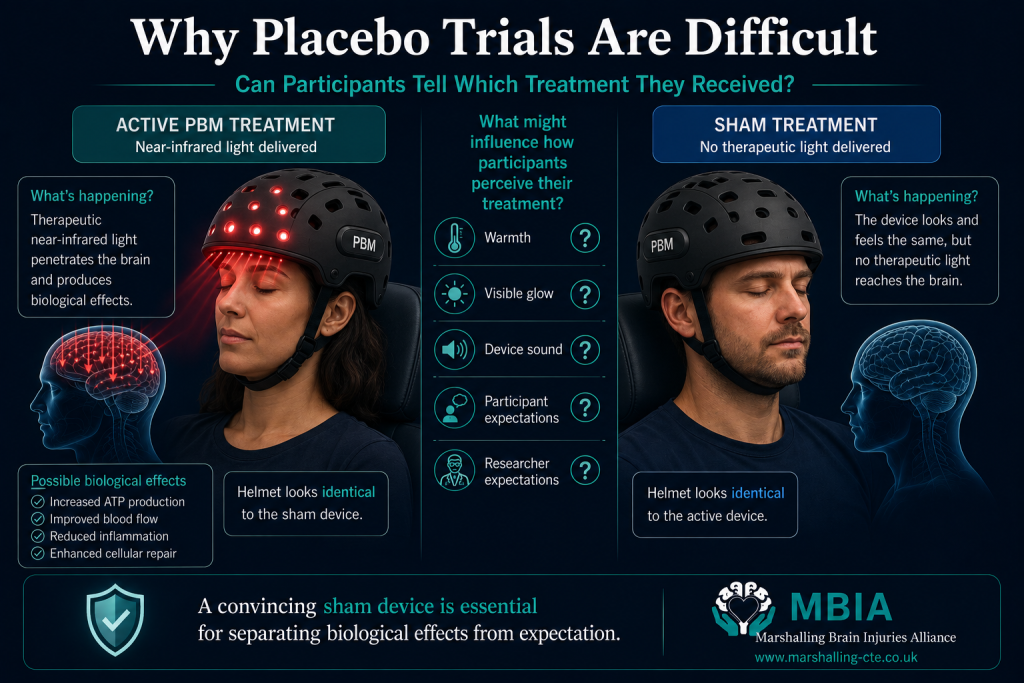
A convincing sham device should:
- Look identical.
- Make the same sounds.
- Feel similar on the head.
- Produce similar warmth.
- Follow the same treatment schedule.
- Avoid delivering a biologically meaningful dose.
If the active device becomes warm or produces visible light, participants may guess which group they are in.
Once someone believes they are receiving the real treatment, their expectations can influence how they report symptoms such as:
- Pain.
- Fatigue.
- Sleep.
- Mood.
- Brain fog.
- Perceived concentration.
Researchers and caregivers may also form expectations if they can identify the active device.
This is known as a blinding problem.
It does not mean reported improvements are imaginary.
It means scientists need especially convincing sham devices and objective outcome measures to separate the biological treatment effect from expectation.
Cognitive Testing Can Improve Through Practice
Many PBM trials use memory, attention or executive-function tests before and after treatment.
These tests are important, but they create another challenge.
People can sometimes improve simply because they have completed the test before.
They may remember the instructions.
They may develop a better strategy.
They may feel less anxious during the second assessment.
This is called a practice effect.
A small increase in a cognitive score does not always mean the brain has biologically improved.
Well-designed trials try to reduce this problem by using:
- Alternative versions of tests.
- Sham comparison groups.
- Longer intervals between assessments.
- Predefined primary outcomes.
- Measures of everyday function as well as cognitive scores.
The most convincing evidence would show not only improved test performance, but also meaningful changes in daily life.
Researchers Measure Many Different Outcomes
Some trials focus on:
- Memory.
- Attention.
- Reaction time.
- Sleep.
- Pain.
- Fatigue.
- Depression.
- Post-concussion symptoms.
- Cerebral blood flow.
- Brain oxygenation.
- MRI connectivity.
- Electrical brain activity.
- Quality of life.
Measuring several outcomes can provide a detailed picture.
It can also create a statistical problem.
If researchers test enough outcomes, one may appear positive simply by chance.
Imagine throwing many darts at a wall.
Even without perfect aim, one may eventually land near the centre.
This is why researchers should identify their main outcome before the study begins and register their analysis plan publicly.
When a study highlights only its most positive result after testing many measures, the finding should be interpreted cautiously.
MRI Changes Are Not the Same as Recovery
Brain-imaging studies have shown changes in white matter, blood flow and functional connectivity after PBM.
These findings are valuable because they suggest that the treatment may be biologically active.
However, an altered scan is not automatically a clinical benefit.
Someone may show a change on MRI without experiencing:
- Better memory.
- Less fatigue.
- Greater independence.
- A successful return to work.
- Improved relationships.
- Better quality of life.
Scientists need to demonstrate that imaging changes are linked with outcomes that matter to patients.
Otherwise, a scan may show target engagement—evidence that the treatment affected the brain—without proving that it improved the person’s life.
Positive Results Are More Likely to Be Published
Another challenge is publication bias.
Studies with exciting or positive findings are more likely to be:
- Submitted to journals.
- Accepted for publication.
- Reported in the media.
- Shared by treatment providers.
- Promoted online.
A study showing no benefit may receive far less attention—or may never be published at all.
This can make a treatment appear more effective than it really is.
Imagine reading reviews for a product when only the happiest customers leave comments.
The available reviews would not represent everyone’s experience.
Systematic reviews try to identify publication bias, but this is difficult when unpublished trial data cannot be accessed.
Trial registration and complete reporting are therefore essential.
Researchers should publish results even when the treatment does not work as hoped.
Negative studies are not failures.
They help identify ineffective doses, unsuitable devices and patient groups that may not benefit.
Industry Funding and Conflicts of Interest
Developing PBM equipment costs money.
Manufacturers may provide devices, technical expertise or research funding.
Some scientists may also help design a device, hold patents, receive consultancy payments or have a financial interest in the company producing it.
Industry involvement does not automatically make a study unreliable.
Many important medicines and medical technologies were developed through collaboration between researchers and companies.
The concern is that financial relationships may influence:
- Study design.
- Which outcomes are selected.
- How results are analysed.
- Which findings are emphasised.
- Whether disappointing results are published.
- The language used in conclusions and marketing.
This is why conflicts of interest must be declared clearly.
The strongest evidence will come from:
- Independent research teams.
- Preregistered study protocols.
- Transparent statistical plans.
- Complete reporting of all outcomes.
- Replication using the same protocol.
- Access to anonymised study data where appropriate.
Trust grows when research can be checked and repeated by people with no financial stake in the result.
Why Larger Trials Are Essential
Larger trials do much more than increase participant numbers.
They help researchers answer questions that small pilots cannot.
A strong future PBM trial should ideally:
Include Enough Participants
Hundreds of participants may be needed to separate a genuine treatment effect from natural variation and chance.
Recruit From Several Centres
A multicentre trial reduces the risk that results depend on one clinic, one research team or one unusually selected group of patients.
Use a Credible Sham Device
Participants, clinicians and outcome assessors should be blinded wherever possible.
Use One Clearly Defined Protocol
The wavelength, power, pulse pattern, treatment area, session length and number of sessions should be reported in enough detail for other researchers to reproduce the study.
Select a Primary Outcome in Advance
Researchers should decide before the trial which result will determine whether the treatment succeeded.
Measure Everyday Function
Trials should assess independence, return to work, communication, daily activities and quality of life—not only laboratory tests or brain scans.
Follow Participants for Longer
A treatment that improves a score for two weeks may not provide a lasting benefit.
Longer follow-up is needed to determine durability and safety.
Include Diverse Participants
Research should reflect differences in age, sex, ethnicity, skin pigmentation, hair type, injury severity, disease stage and other medical conditions.
Report All Results
Positive, negative and inconclusive findings should all be published.
What Would Convincing Evidence Look Like?
For brain injury, convincing evidence would show that people receiving PBM experience better recovery than those receiving sham treatment in areas such as:
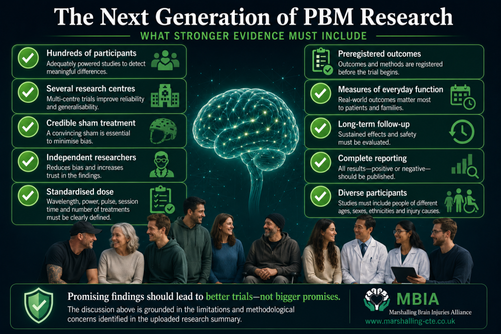
- Cognition.
- Symptom burden.
- Independence.
- Return to work or education.
- Quality of life.
- Long-term neurological function.
For Alzheimer’s disease or mild cognitive impairment, convincing evidence would need to show:
- Sustained cognitive benefit.
- Better daily functioning.
- Slower progression over time.
- Benefits beyond practice effects.
- Acceptable long-term safety.
- Independent replication.
Biomarker or MRI changes would strengthen the case—but they would not be enough on their own.
The treatment must ultimately make a meaningful difference to patients and families.
What We Know
🟢 PBM produces measurable biological effects in cells, animals and some human studies.
🟢 Small trials have reported encouraging changes in cognition, symptoms, blood flow and brain imaging.
🟢 Short-term treatment has generally appeared well tolerated in carefully selected research participants.
🟢 The field has progressed from case reports towards randomised, sham-controlled trials.
What We Don’t Yet Know
🟠 Which device and wavelength are most effective.
🟠 What dose should reach the scalp—and the brain.
🟠 How often treatment should be given.
🟠 Which patients are most likely to respond.
🟠 Whether benefits remain after treatment stops.
🟠 Whether scan changes reliably predict real-life recovery.
🟠 Whether positive findings will be reproduced independently.
🟠 Whether long-term repeated treatment is safe.
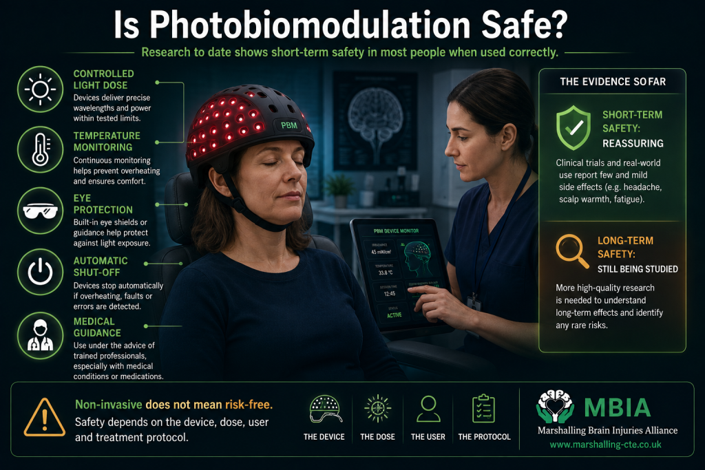
Early studies are reassuring—but “non-invasive” does not mean completely risk-free.
Safety is one of the first questions anyone should ask before trying a new treatment.
Photobiomodulation is often described as non-invasive, because it does not involve surgery, injections or ionising radiation. The red or near-infrared light used in PBM is delivered at intensities intended to influence cellular activity without cutting, burning or destroying tissue.
That makes it very different from surgical lasers and radiotherapy.
Across the small human studies conducted so far, transcranial photobiomodulation has generally appeared to be well tolerated over the short term.
However, this needs to be interpreted carefully.
Most studies have included relatively small numbers of carefully selected participants and have followed them for weeks or months—not years. Researchers therefore know much more about the immediate tolerability of PBM than they do about the effects of repeated, long-term use.
The most accurate conclusion is:
Short-term research findings are broadly reassuring, but long-term safety is not yet fully established.
What Have Human Studies Reported?
In many brain-PBM studies, participants completed treatment without experiencing serious problems.
Reported side effects have generally been mild and temporary.
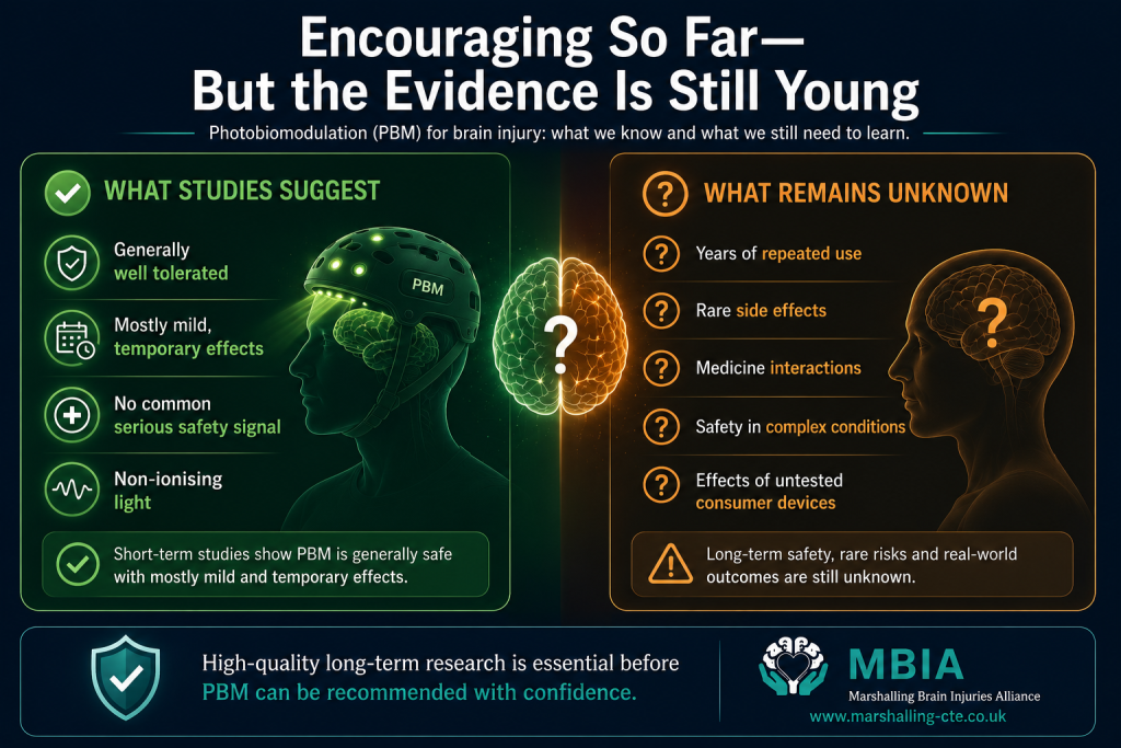
These have included:
- Headache.
- Scalp warmth.
- Mild skin discomfort.
- Tiredness.
- Temporary changes in sleep.
- Dizziness or feeling light-headed.
- Sensitivity to the device or treatment environment.
Importantly, similar symptoms have sometimes been reported by people using sham devices, making it difficult to determine whether they were caused by the light itself, the pressure or warmth of the equipment, participant expectations or the neurological condition being studied.
In the 2020 randomised study of people with acute moderate traumatic brain injury, researchers reported no treatment-related adverse events.
Early dementia and mild cognitive impairment studies have also generally described short-term treatment as reasonably well tolerated.
This is encouraging.
But it is not the same as proving that every PBM device, dose or treatment schedule is safe for everyone.
Why “Non-Thermal” Does Not Mean No Heat at All
Photobiomodulation is often described as a non-thermal treatment.
This means the intended biological effect does not depend on heating or destroying tissue.
However, any powered light device can produce some warmth.
The amount depends on:
- The power of the light source.
- How closely the device touches the scalp.
- Whether heat can escape.
- The length of the session.
- The number and arrangement of LEDs.
- The condition and quality of the device.
- The surrounding temperature.
A properly designed research device should control heat and remain within safe operating limits.
A poorly manufactured or incorrectly used consumer device may not.
This distinction matters because the word “low-level” can create the impression that a device is automatically incapable of causing discomfort or excessive heating.
Device design, maintenance and correct use remain important.
Eye Safety Matters
Red and near-infrared light may appear gentle, but intense light should not be directed into the eyes unless a device has been specifically designed and tested for that purpose.
Near-infrared light is particularly important because it may be invisible.
A person cannot rely on discomfort or brightness alone to judge whether their eyes are receiving too much exposure.
Eye-safety requirements depend on:
- Wavelength.
- Optical power.
- Distance from the eye.
- Exposure time.
- Whether the beam is focused or widely dispersed.
- The design of the device.
Some systems may require protective eyewear.
Others may be enclosed so that light cannot reach the eyes directly.
Users should follow the instructions for the specific device rather than assuming that all red or near-infrared products require the same precautions.
What About Photosensitive Conditions and Medicines?
Some medical conditions and medicines can make the skin or eyes more sensitive to light.
These are often described as photosensitising.
Examples can include certain:
- Antibiotics.
- Acne medicines.
- Diuretics.
- Antidepressants.
- Antipsychotic medicines.
- Anti-inflammatory medicines.
- Cancer treatments.
- Herbal products.
The degree of risk depends on the medicine, the wavelength used and the person’s individual health.
This does not mean that everyone taking one of these medicines will experience a problem.
It means that people should not assume PBM is automatically suitable simply because it is sold as a wellness treatment.
Anyone using photosensitising medication—or living with a condition that affects light sensitivity—should seek advice from an appropriate healthcare professional before using a high-output device.
People should not stop prescribed medication in order to use photobiomodulation.
Long-Term Safety Remains Uncertain
One of the biggest gaps in the evidence is long-term exposure.
Many commercial protocols recommend repeated treatment several times per week, sometimes indefinitely.
Yet most clinical studies have lasted only a few weeks or months.
Researchers do not yet have enough high-quality evidence to answer questions such as:
- Is daily use safe over several years?
- Could repeated exposure produce effects that are not visible in short trials?
- Is lifelong maintenance treatment necessary?
- Does long-term safety differ between wavelengths?
- Are some brain conditions more sensitive than others?
- Could treatment interact with neurological medicines?
- Does the risk change when several light devices are used together?
So far, studies have not identified a clear major safety signal.
However, absence of evidence is not the same as evidence of absence.
Rare side effects may not appear until thousands of people have been treated and followed for longer periods.
This is one reason larger clinical trials and post-market monitoring are important.
Could PBM Stimulate Something We Don’t Want to Stimulate?
Because PBM is intended to influence cellular metabolism, some people understandably worry about whether it could stimulate abnormal cells.
This is a complicated area.
Photobiomodulation is already used in some supportive cancer-care settings—for example, to reduce oral mucositis caused by treatment—and professional protocols distinguish supportive PBM from attempts to treat a tumour directly.
However, the safety of applying light over a known or suspected tumour depends on the clinical situation, treatment area, dose and medical advice.
Someone with:
- A known brain tumour.
- An unexplained mass.
- Active cancer in the intended treatment area.
- New neurological symptoms that have not been investigated.
should not rely on a consumer device or online advice.
They should discuss treatment with the medical team responsible for their care.
The same principle applies to any serious or unexplained neurological symptoms: PBM should never delay appropriate diagnosis.
Regulation: What Does “Cleared” or “Registered” Really Mean?
Regulation is one of the most confusing parts of the PBM market.
A company may advertise that its product is:
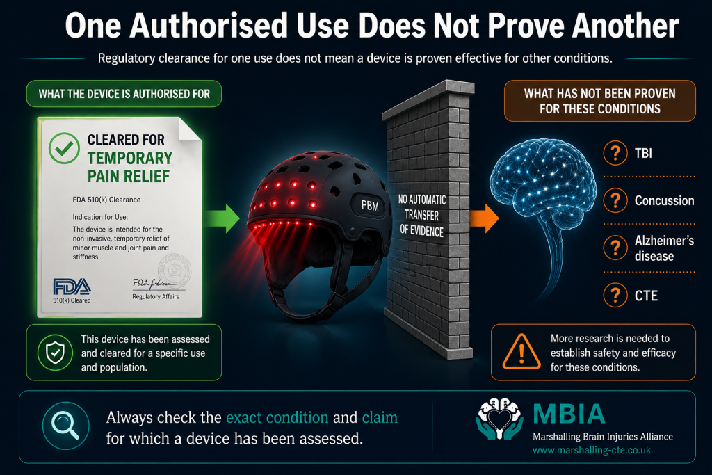
- Registered.
- Certified.
- CE marked.
- UKCA marked.
- FDA cleared.
- A medical device.
- Clinically tested.
These phrases do not all mean the same thing.
More importantly, they do not necessarily mean the device has been authorised to treat traumatic brain injury, concussion, Alzheimer’s disease or another neurological condition.
A device may have regulatory clearance for a very different purpose, such as:
- Temporary pain relief.
- Increased local circulation.
- Muscle relaxation.
- Cosmetic skin treatment.
- General wellness.
That clearance cannot automatically be transferred to brain injury or dementia.
Think of it like a medicine approved to treat one condition.
The fact that it has passed regulatory checks for that use does not mean it has been proved effective for every other condition.
Consumers should therefore ask:
What exact claim has the device been assessed or authorised for?
Not simply:
Does the company use a regulatory logo?
A Medical Device Is Not the Same as a Proven Treatment
Medical-device regulation often focuses on whether the equipment meets standards relating to manufacture, electrical safety, performance and the claims made by the company.
Clinical effectiveness for a particular disease is a separate question.
A helmet may safely deliver near-infrared light at the output described by the manufacturer.
That does not prove it improves memory after concussion.
An intranasal device may operate within electrical and optical safety standards.
That does not prove it slows Alzheimer’s disease.
Safety, technical performance and clinical effectiveness are all important—but they are not interchangeable.
The Consumer Red-Light Market Is Extremely Varied
The growth of red-light therapy has created a huge consumer market.
Products now include:
- Full-body panels.
- Face masks.
- Handheld devices.
- Headbands.
- Caps.
- Helmets.
- Intranasal applicators.
- Low-cost devices sold through online marketplaces.
Some are produced by established manufacturers and provide detailed technical information.
Others make broad health claims while providing little evidence about what the product actually delivers.
A device may advertise a wavelength such as 810 or 850 nanometres but fail to clearly explain:
- Its measured optical output.
- The dose reaching the treatment area.
- Whether the light is continuous or pulsed.
- The pulse frequency and duty cycle.
- The size of the treatment area.
- How heat is controlled.
- Whether output has been independently verified.
- Which clinical study used that exact device and protocol.
Without this information, it is difficult to compare the product with published research.
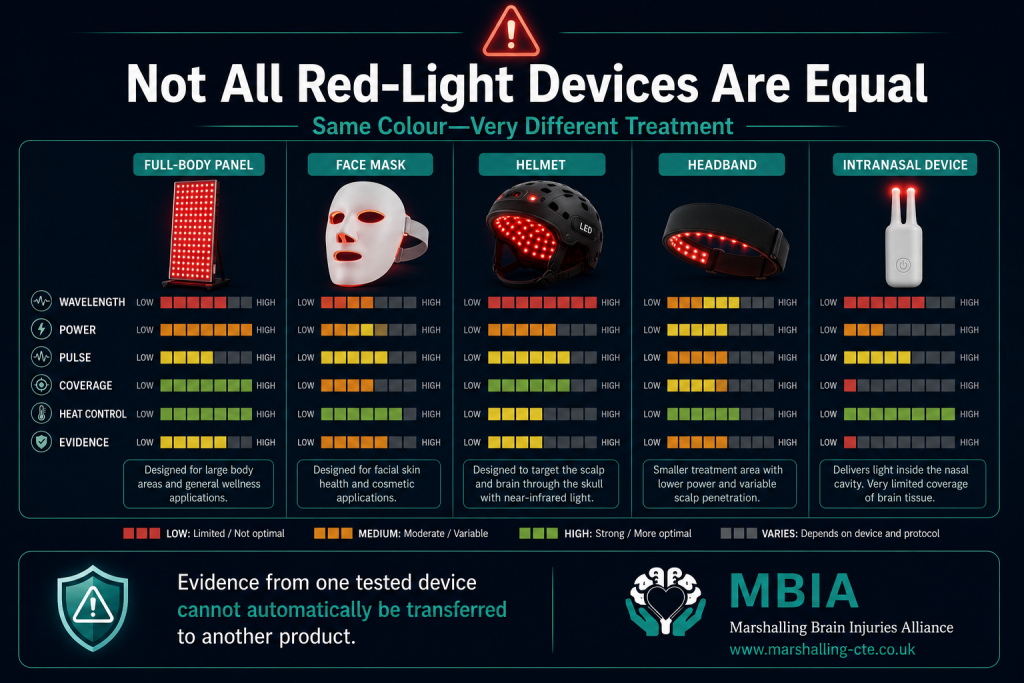
Two products can both be advertised as “near-infrared brain devices” while delivering completely different treatments.
One may contain a carefully arranged array of LEDs in direct contact with the scalp.
Another may use a few weak lights positioned several centimetres away.
One may deliver a measured research dose.
Another may provide too little energy to create the same biological effect.
A third may deliver far more than was used in a clinical study.
Even products using the same wavelength may differ in:
- Power.
- Beam shape.
- Pulse pattern.
- Coverage.
- Treatment time.
- Heat production.
- Manufacturing quality.
- Electrical safety.
- Accuracy of the stated specifications.
This is why evidence from one studied device cannot automatically be used to advertise another.
The relevant intervention is not simply “red light.”
It is the exact wavelength, dose, device design, placement and schedule tested in the study.
Be Cautious of Devices That Promise Too Much
Warning signs include claims that a device:
- Repairs brain damage.
- Reverses dementia.
- Clears amyloid or tau.
- Cures persistent concussion symptoms.
- Prevents CTE.
- Works for everyone.
- Has no risks or side effects.
- Is proven because it uses the same wavelength as a study.
- Replaces medication or rehabilitation.
These claims go beyond the current human evidence.
A responsible company should clearly separate:
- Laboratory findings.
- Animal research.
- Early human trials.
- Regulatory status.
- Proven clinical indications.
- Conditions still being investigated.
Promotional testimonials can be emotionally powerful, but they cannot show whether improvements resulted from the device, natural recovery, rehabilitation, expectation or normal changes in symptoms.
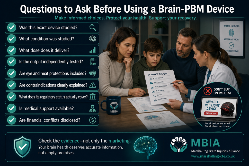
Before purchasing a consumer device, consider asking:
Has This Exact Device Been Studied?
Not merely a similar device using a similar wavelength.
What Condition Was Studied?
Research for pain or skin health does not prove benefit for the brain.
Was the Study Randomised and Sham Controlled?
Case reports and testimonials provide much weaker evidence.
What Dose Does the Device Deliver?
Look for wavelength, irradiance, fluence, pulse pattern and session duration.
Has the Output Been Independently Tested?
Manufacturer specifications are most useful when verified.
What Safety Features Are Included?
This may include heat control, automatic shut-off, eye protection and electrical-safety certification.
Are Contraindications Clearly Listed?
A responsible manufacturer should explain when users need medical advice.
What Does the Regulatory Claim Actually Cover?
Check the specific intended use, not only the presence of a logo or registration number.
Are Conflicts of Interest Declared?
Find out whether the research was funded or conducted by the manufacturer.
Is There Ongoing Clinical Support?
Brain injury and dementia are complex conditions. A product should not be marketed as a substitute for appropriate medical assessment.
PBM Should Not Replace Established Care
Perhaps the greatest safety risk is not always the light itself.
It may be what someone gives up—or delays—because they believe a device will treat the underlying problem.
Photobiomodulation should not be used as a reason to avoid:
- Emergency assessment after a serious head injury.
- Investigation of worsening neurological symptoms.
- Prescribed Alzheimer’s medication.
- Concussion management.
- Rehabilitation.
- Psychological support.
- Treatment for sleep disorders.
- Management of epilepsy.
- Support for depression or suicidal thoughts.
- Medical investigation of headaches, weakness or personality changes.
PBM may eventually become a useful addition to neurological care.
It should not be presented as a replacement for diagnosis, rehabilitation or evidence-based treatment.
What We Know
🟢 Short-term PBM has generally appeared well tolerated in small human studies.
🟢 Serious treatment-related adverse events have not emerged as a common finding in the published brain-PBM trials so far.
🟢 PBM uses non-ionising light and is fundamentally different from radiotherapy.
🟢 Properly designed PBM is intended to create photochemical effects rather than burn or destroy tissue.
🟢 Device design, optical output, heat management and eye safety all matter.
What We Don’t Yet Know
🟠 Whether frequent PBM use remains safe over many years.
🟠 Whether rare side effects will appear in much larger populations.
🟠 How PBM interacts with every medicine and neurological condition.
🟠 Whether long-term safety differs between devices and doses.
🟠 Whether some patient groups require additional precautions.
🟠 Whether consumer-device specifications consistently match their advertised output.
🟠 What level of medical oversight is appropriate for home brain-PBM treatment.
The MBIA View
Photobiomodulation has a reassuring short-term safety record in the small clinical trials completed so far.
That is important.
However, “appears safe in early studies” is not the same as “proven safe for everyone, at every dose, indefinitely.”
At MBIA, we believe people should be able to explore emerging therapies without being misled by vague regulatory language or exaggerated safety claims.
Anyone considering PBM for brain injury or cognitive impairment should look beyond the colour of the light and examine the complete device, dose, evidence and intended use.
We also believe companies have a responsibility to be transparent.
They should clearly explain:
- What their device has been tested for.
- Which claims are supported by human trials.
- What remains experimental.
- Who should seek medical advice.
- What financial relationships exist within the research.
Hope should never depend on hiding uncertainty.
MBIA View
Photobiomodulation is one of the most scientifically plausible emerging therapies in neuroscience. Unlike many treatments promoted online, there is a genuine biological rationale supported by decades of laboratory research and an increasing number of human clinical trials.
However, promising biology does not automatically become proven medicine.
At MBIA we believe people living with brain injury deserve evidence-based hope—not exaggerated claims or false promises. The research into photobiomodulation is advancing rapidly, and while the results are encouraging, larger high-quality studies are still needed before it can become part of routine neurological care.