Why Does This Happen?
To understand why, it helps to understand what different brain scans can and cannot show.
No scan is perfect.
Each type of scan is designed to look at different things.
Some scans are very good at identifying structural damage.
Others are designed to look at brain function, metabolism, blood flow, or connectivity.
What Is an MRI?
MRI stands for:
Magnetic Resonance Imaging
MRI uses powerful magnets and radio waves to create detailed pictures of the brain.
It is excellent at identifying:
- Tumours
- Bleeding
- Stroke
- Structural abnormalities
- Brain shrinkage (atrophy)
- Significant areas of damage
MRI is one of the most useful imaging tools available in modern medicine.
However, it has limitations.
Why Concussions Often Don't Show Up on MRI
Most concussions are considered functional injuries rather than large structural injuries.
Following a concussion, there may be:
- Microscopic damage
- Changes in brain connectivity
- Neurochemical changes
- Metabolic changes
- Inflammation
These changes are often too small to be detected on routine MRI scans.
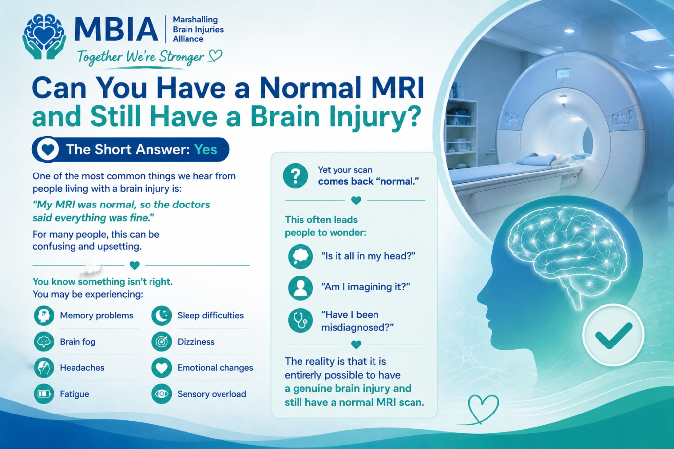
This is why many people with genuine concussion symptoms have completely normal imaging.
A Normal MRI Does Not Mean a Normal Brain
This is one of the most important things to understand.
A standard MRI shows the structure of the brain.
It does not always show how the brain is functioning.
Think of it like this:
A mechanic can look at the outside of a car and see that it appears intact.
That does not mean the engine is running properly.
Similarly, a brain may appear structurally normal while still experiencing functional problems.
The Difference Between Structural & Functional Problems
Structural Problems
Things that can often be seen on standard imaging:
- Bleeding
- Swelling
- Skull fractures
- Large lesions
- Significant tissue damage
Functional Problems
Things that may not appear on routine imaging:
- Brain fog
- Memory difficulties
- Fatigue
- Concentration problems
- Emotional regulation difficulties
- Sensory overload
A person may experience severe symptoms despite having no obvious abnormalities on standard scans.
What About Repeated Head Impacts?
Repeated head impacts can sometimes affect:
- White matter pathways
- Brain connectivity
- Neural communication
These changes may not always be visible on routine MRI.
This is one reason why athletes, military personnel, domestic abuse survivors, and others with repeated head trauma may experience symptoms despite "normal" scans.
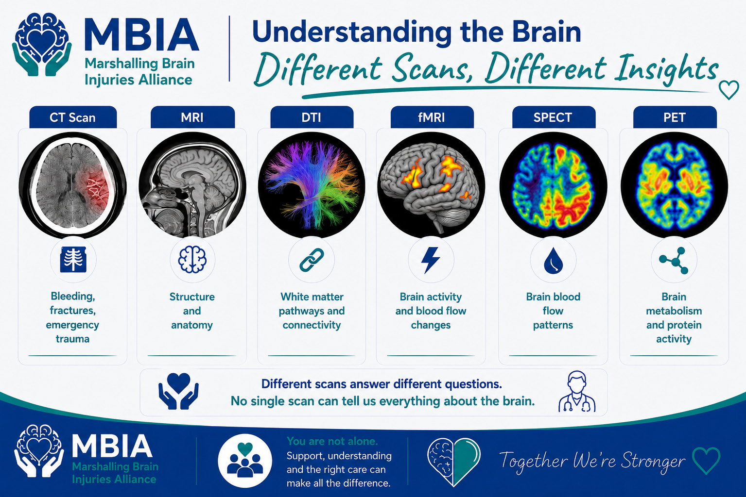
CT Scan
Best For:
- Emergency assessment
- Bleeding
- Skull fractures
- Major trauma
Limitations:
May miss concussion and subtle brain injury changes.
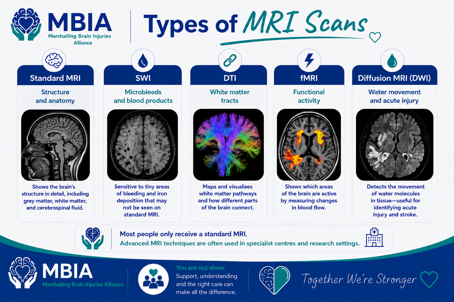
Standard MRI
Best For:
- Structural abnormalities
- Tumours
- Stroke
- Significant injury
Limitations:
Often normal in concussion and many mild brain injuries.
Advanced MRI Techniques
Some MRI techniques can provide more detailed information about brain structure and connectivity.
Examples include:
- DTI (Diffusion Tensor Imaging)
- SWI (Susceptibility Weighted Imaging)
- Functional MRI (fMRI)
These are often used in research settings and specialist clinics
DTI (Diffusion Tensor Imaging)
DTI examines white matter pathways within the brain.
White matter acts like the brain's communication network.
DTI may identify abnormalities in brain connectivity that are not visible on standard MRI.
However:
- It is not routinely used in most healthcare systems.
- It is not currently considered a standalone diagnostic tool for concussion or CTE.
Functional MRI (fMRI)
fMRI looks at brain activity by measuring changes in blood flow.
It helps researchers understand how different areas of the brain function.
It is primarily used in research and specialist settings.
SPECT Imaging
SPECT looks at blood flow patterns within the brain.
It may identify areas receiving reduced blood flow.
Some clinics use SPECT to explore functional brain changes.
However, its role remains an area of ongoing research and debate.
PET Imaging
PET scans examine brain metabolism and biological activity.
In research settings, specialised PET tracers are being used to investigate conditions such as:
- Alzheimer's disease
- Tau pathology
- Chronic Traumatic Encephalopathy (CTE)
At present, PET imaging for CTE remains primarily a research tool.
Different Types of Brain Imaging
Why Symptoms Matter More Than Scans
One of the biggest mistakes people make is assuming:
"If the scan is normal, nothing is wrong."
Brain injury diagnosis is not based on scans alone.
Healthcare professionals also consider:
- Symptoms
- Medical history
- Mechanism of injury
- Cognitive testing
- Neurological examination
A person with severe symptoms and a normal MRI still deserves support, treatment, and understanding.
The Emotional Impact of a Normal Scan
Many people describe mixed emotions when told their scan is normal.
At first they feel relieved.
Then confusion sets in.
Questions often follow:
"Why do I still feel so awful?"
"Why can't anyone explain my symptoms?"
"Why does nobody believe me?"
These feelings are understandable.
A normal scan does not invalidate your symptoms.
A Message for Patients
If you have ongoing symptoms but a normal MRI:
Please remember:
Your symptoms are real.
A normal scan does not automatically mean nothing is wrong.
Many people living with concussion, PCS, TBI, and other neurological conditions have normal imaging.
Recovery and treatment should focus on the whole person, not just the scan result.
A Message for Families
A normal scan can sometimes make it harder for family members to understand what their loved one is experiencing.
It is important to remember that many brain injury symptoms are invisible.
Just because damage cannot be seen on a routine scan does not mean the person is not struggling.
Understanding this can make a huge difference.
Key Takeaways
- Yes, it is possible to have a brain injury despite having a normal MRI.
- Standard MRI scans are designed to identify structural abnormalities.
- Many concussion-related changes occur at a microscopic or functional level and may not appear on routine imaging.
- Different scans provide different types of information.
- Symptoms, history, and clinical assessment are often more important than imaging alone.
- A normal scan does not mean symptoms are imagined or unimportant.
- Research into advanced imaging and biomarkers continues to improve understanding of brain injury.
Continue Exploring
Together We're Stronger